Tumors in female dog anatomy are described as new growths that are abnormal by nature and can be further defined as benign or malignant. Tumors possess the ability of uncontrolled and rapid cellular proliferation.
A benign tumor is a type of tumor that does not invade the nearby tissue and does not metastasize to other organs. Malignant tumors have the ability to spread from the origin to virtually every part of the body where they continue to grow and form new tumors at that site.
In canines, the reproductive system is not spared when it comes to tumors. Every part of the reproductive system, both in male and female dogs is prone to cancer development.

Mammary Tumors
These tumors are most frequent in intact female dogs, and extremely rare in male dogs. Most often, the two posterior mammary glands are involved as opposed to the three anterior glands.
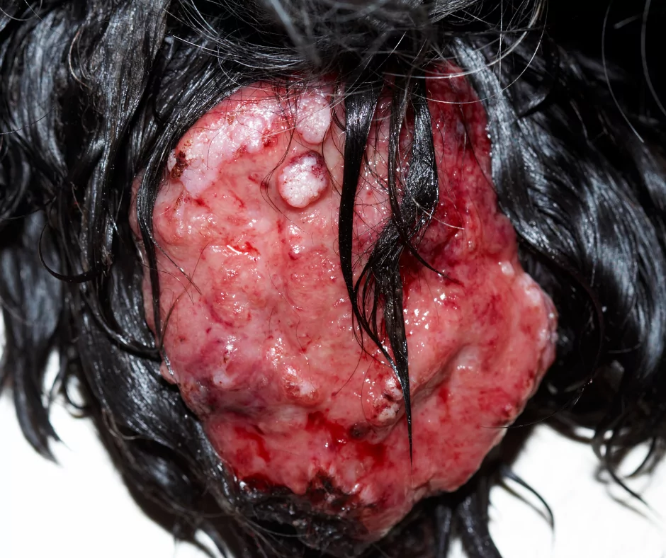
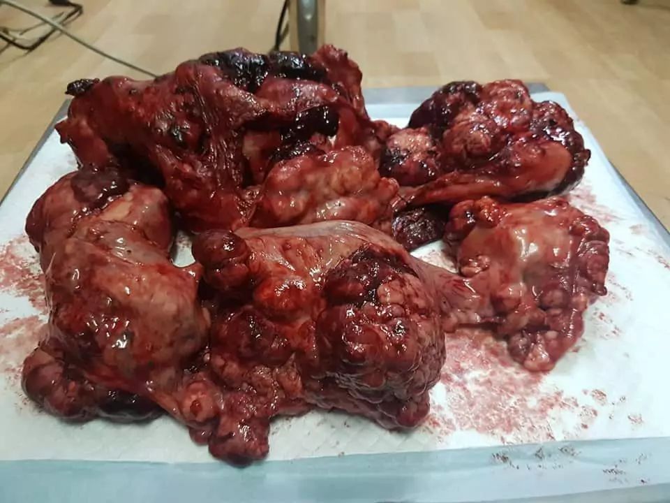
These tumors appear as single or multiple modules (1-25cm) in one or more glands. The surface of the nodule is usually lobulated, grey in color, firm, and sometimes contains cysts filled with fluid. There can be mixed mammary tumors that can contain cartilage or bone on the cut surface.
Ovariectomy generally decreases the risk of developing mammary neoplasia. Spaying before the first estrus lowers the risk of mammary tumors to 0.5%, and after the first estrus to 8%. Female dogs that are spayed after maturity are considered at the same level of risk of developing mammary tumors as intact female dogs.
The most common indication of a mammary tumor is a palpable mass under the skin in the mammary area. Other signs may include discharge from the mammary gland, ulceration of the skin over the gland, swelling of the mammary gland, pain, inappetence, loss of weight, and weakness.
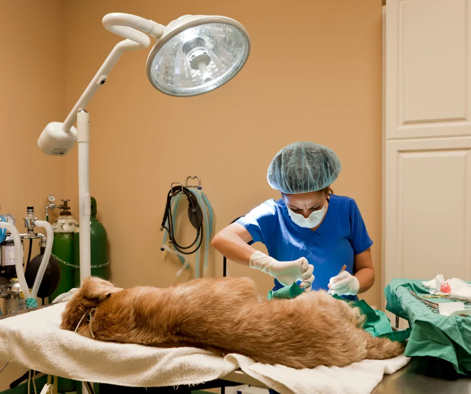
Mammary tumors are almost always treated surgically. Options are the following: lumpectomy (removal only of the tumor), simple mastectomy (removal only of the affected gland), modified radical mastectomy (removal of the tumorous gland along with the glands that share the same lymphatic drainage and the associated lymph nodes), and radical mastectomy (removal of the whole mammary chain along with the associated lymph nodes).
Till now, chemotherapy has not shown as an effective treatment for mammary tumors in dogs.

Ovarian Tumors
Ovarian tumors are very rare in dogs. When they develop, most often arise between four to 15 years of age. There are documented ovarian tumors in much younger dogs.
These tumors are divided into categories based on their cell origin. They can be epithelial tumors, germ cell tumors, and sex cord-stromal cell tumors.

Usually, these tumors do not show symptoms unless they grow too big. When they grow too big, they may produce a buildup of carcinogenic fluid in the abdomen, extra fluid between the layers of the pleura outside the lungs, or hormonal dysfunction.
When an increase of estrogen and progesterone happens, it results in the enlarging of the vulva, blood-like discharge, prolonged estrus, alopecia, and a low RBC count.
Treatment of ovarian tumors is always complete ovariohysterectomy and chemotherapy.
Uterine Tube Tumors in Female Dog Anatomy
The occurrence of tumors in the uterine tube is very rare. These tumors are usually of mesenchymal origin. Lipomas are rare tumors of the uterine tube and are located on the ovarian bursa. The mass has the characteristics of mature adipose tissue. Also, adenocarcinomas have been reported in dogs.
Uterine Tumors
These tumors in dogs are most often benign and very rare. Usually, they develop in dogs that are middle-aged to older that obviously have not been spayed. These tumors form from the uterine smooth muscle and epithelial tissue. Around 90% of ovarian tumors are diagnosed as leiomyomas (benign). A small percentage of dogs are unfortunate to develop the malignant form–leiomyosarcoma.
Most often there are no signs of these tumors, but they may show vaginal discharge, pyometra, and infertility. Treatment is always surgical removal of the tumors with a complete ovariohysterectomy.
Vaginal and Vulvar Tumors in Female Dog Anatomy

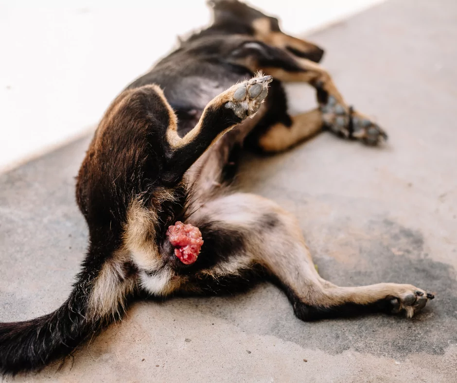
These tumors are the second most common tumors that can develop in female dogs after the mammary gland tumors. Unlike mammary tumors, vaginal lesions are benign, non-cancerous, and originate from the smooth muscle tissues.
The most common type of vaginal and vulvar tumors reported in dogs are leiomyomas, fibroleiomyomas, fibromas, polyps, lipomas, adenomas, myxomas, myxofibromas, and others.
There are also malignant tumors as well, like the TVT (transmissible venereal tumor), adenocarcinomas, hemangiosarcoma, and the mast cell tumor. The most susceptible are unsprayed dogs that have never given birth between the ages of two and 18 years.
Symptoms of vaginal or vulvar tumors may include vulvar bleeding or discharge, vulvar mass, dysuria, hematuria, tenesmus, excessive licking at the vulvar area, and dystocia. Because it has been found that vaginal and vulvar tumors are hormone-dependent, the treatment of choice is a complete ovariohysterectomy.
This procedure allows visual examinations of the abdominal organs for possible metastasis. Conservative surgical extirpation combined with ovariohysterectomy will result in complete remission of benign tumors.
