
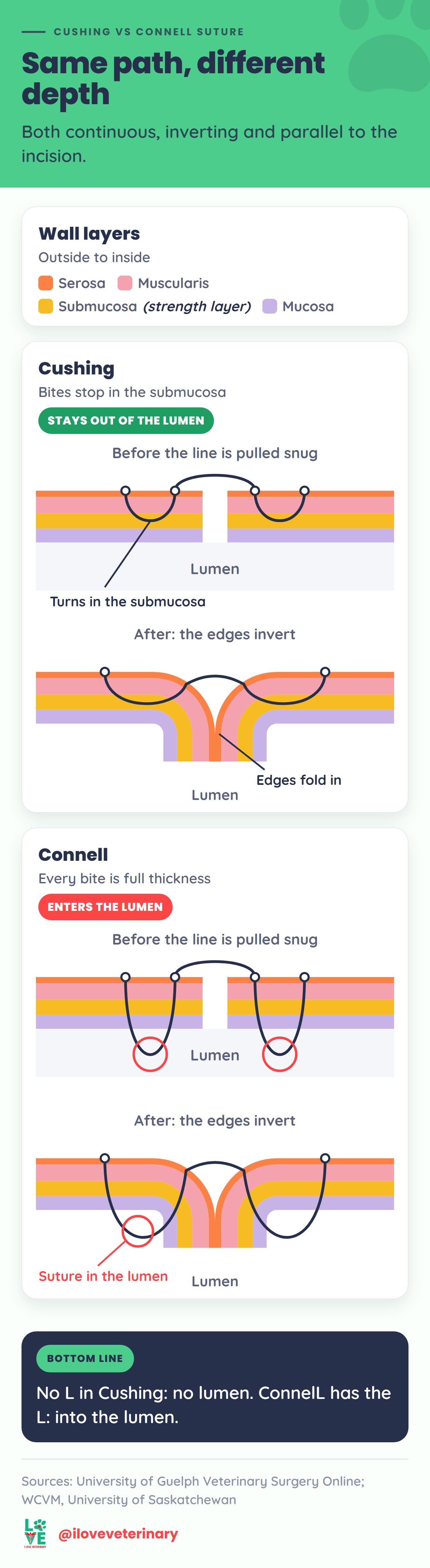
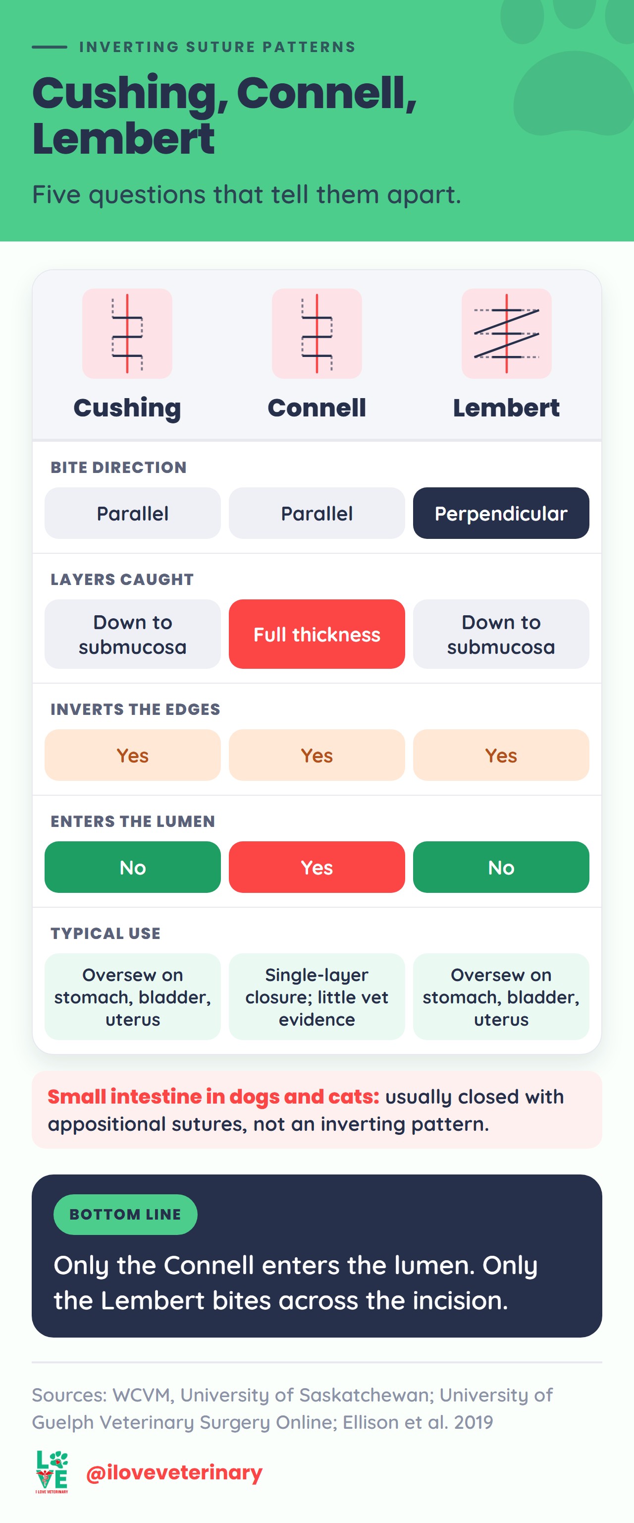
Cushing and Connell are close cousins. Both are continuous, inverting suture patterns for hollow organs such as the stomach, urinary bladder and uterus, and both run their bites parallel to the incision. The one difference that matters: the Connell passes through the full wall into the lumen, while the Cushing stops in the submucosa and stays out of it.
The memory trick from the University of Saskatchewan's surgery lab: Connell has the "L", so it goes into the lumen. Cushing has no "l", so no lumen (WCVM).

Cushing vs Connell at a glance
Pattern | Bite direction | Layers caught | Enters the lumen? | Memory hook |
|---|---|---|---|---|
Cushing | Parallel to the incision | Serosa, muscularis, submucosa | No | No "L" in Cushing: no lumen |
Connell | Parallel to the incision | Full thickness | Yes | ConnelL has the L: it goes into the lumen |
Lembert | Perpendicular to the incision | Serosa, muscularis, submucosa | No | LemberT ends in T, for transverse |
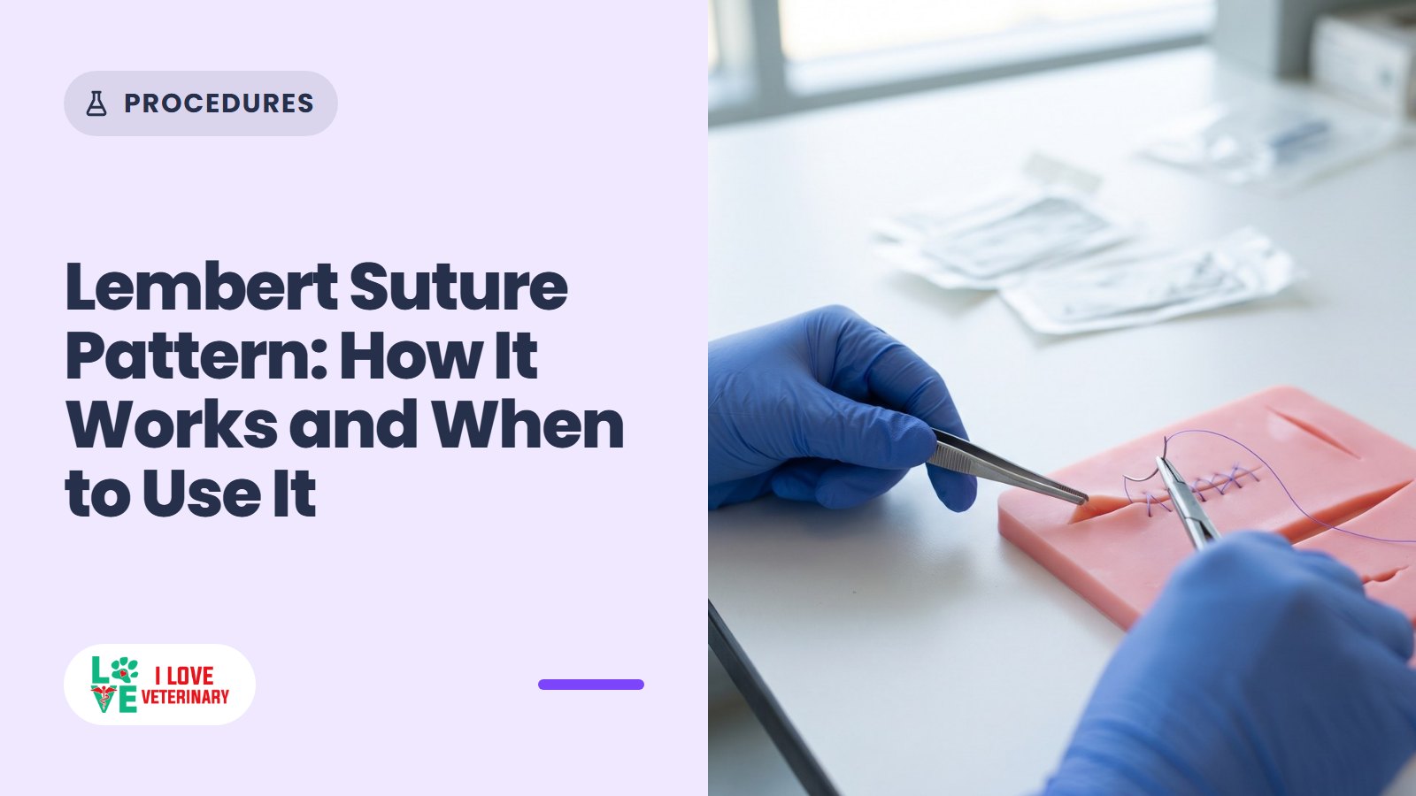
Everything else is shared. They are continuous, so there are knots only at the start and the end of the line. They invert, so the cut edges roll inward. And the farther from the edge the bites are placed, the more tissue folds in. The Lembert is the third inverting pattern students meet in the same lab; its bites cross the incision instead of running along it, and our Lembert suture pattern guide walks through it step by step.
The wall layers both patterns are built around
A hollow organ wall has four layers. From the outside in: the serosa, the muscularis, the submucosa and the mucosa, which lines the lumen (the open space inside the organ). The submucosa is the strength layer, and the University of Guelph's surgery text says it must be included in the bites of an intestinal closure.

That is where the two patterns part ways. A Cushing bite goes through the serosa and muscularis and into the submucosa, then stops. A Connell bite keeps going, through the mucosa and into the lumen.
How the Cushing pattern is placed
Guelph's Veterinary Surgery Online describes the continuous Cushing in this order (VSO, Cushing pattern):
- A partial-thickness simple throw across one end of the incision, tied.
- The needle crosses the incision and goes down into the muscular and submucosal layers, running parallel to the incision. In Guelph's example each bite is about 5-8 mm long.
- The needle comes out on that same side.
- It crosses to the opposite side for the same parallel bite there.
- The pattern repeats to the end of the incision and is knotted.
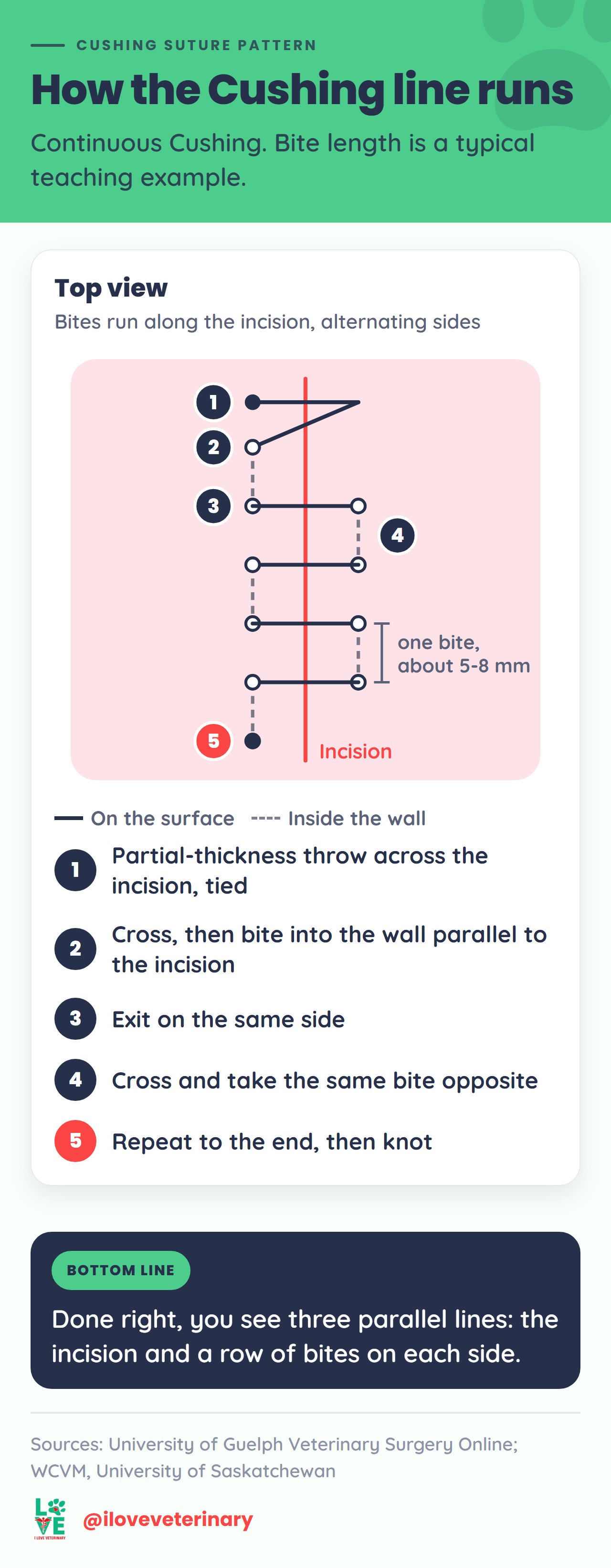
Each pass rolls the cut edges inward as the line is snugged up. Laid over a first layer of sutures, a well-placed Cushing inverts enough tissue to cover that first layer completely, so only its own knots remain visible.
Treat the 5-8 mm as a typical teaching figure, not a rule: bite length scales with the organ. Two of WCVM's rules apply to every bite. The farther from the edge, the more tissue inverts. And tension is set as you go, because it is hard to adjust once the line is finished.
Cushing-style bites also turn up inside another pattern. Guelph's purse-string suture is a ring of partial-thickness, Cushing-type bites around the stump of a hollow organ or around a tube entering one, pulled tight and knotted.
What is the Connell suture technique?
The Connell follows the same path as the Cushing: continuous, parallel bites that cross from side to side and invert the edges. The difference is depth. Every Connell bite passes through the full thickness of the wall, mucosa included, so the needle enters the lumen on each pass (WCVM).
So part of every Connell stitch sits inside the organ, in contact with whatever the organ holds. A Cushing keeps all of its suture in the wall. So when the goal is to keep suture out of the organ's contents, the Cushing (or the Lembert) is the one to reach for.
Going deeper does not make the Connell invert more tissue. With both patterns, inversion comes from how far from the edge the bites are placed.
The name comes from Milwaukee. A 2023 history in The American Surgeon traces the Connell stitch to Milwaukee County Hospital in 1887 and to two generations of surgeons, Dr. M.E. Connell and Dr. F. Gregory Connell (Lundberg and colleagues).
What inverting does, and what it costs
WCVM lists the trade-offs of inverting patterns plainly (suture pattern properties):
- Better seal. Edges turned inward seal better against fluid leaking out of the organ.
- Less exposed suture. A buried suture line means fewer adhesions. University of Minnesota large animal surgery notes make the same point for horses and cattle, and add that small animals form adhesions less readily (UMN).
- A smaller lumen. Every fold that turns in takes up room inside the organ, so inverting patterns are avoided where the lumen is already small.
- Not for skin. Inverted skin edges heal more slowly.
Blood supply is the other cost. Both the Cushing and the Connell have more effect on the local blood supply than a simple continuous line. Overtightening any pattern can cut off the circulation a wound needs to heal, and vessels usually enter the tissue at right angles to the incision. Another reason to set tension bite by bite instead of cinching the whole line at the end.
Where each pattern is used
Dogs and cats: Cushing as the second layer
In small animal surgery the Cushing usually shows up as the oversew: a second layer that inverts and buries a first closure. Guelph's surgery text uses it this way in three common procedures:
- Gastrotomy (an incision into the stomach, often to remove a foreign body like those behind an intestinal blockage in dogs): a full-thickness simple continuous layer, then a Lembert or Cushing oversew. Two inverting lines are another option.
- Cystotomy (an incision into the bladder): a simple continuous appositional layer, then a Lembert or Cushing over the seromuscular layer. A single continuous layer is an alternative, preferred when the bladder wall is thick.
- Hysterotomy during a C-section: a simple continuous layer, sometimes followed by a Cushing or Lembert.
In all three, the inverting oversew is a Cushing or a Lembert. The Connell does not appear in that role in the Guelph text.
A Cushing can also stand alone. In a 2006 study at the University of Florida, 40 rats with experimentally inflamed bladders had their cystotomies closed with either a single-layer continuous Cushing or a simple continuous appositional line. Leak pressure, strength and inflammation did not differ. A rat model shows both closures can work; it does not pick one for a dog.
The small intestine is the big exception. In dogs and cats it is normally closed with appositional sutures, which bring the edges together without folding them in. A 2019 review in Veterinary Surgery explains why: appositional patterns preserve the diameter of the lumen, heal well and match inverting patterns for bursting strength after the first 24 hours. Fold tissue into a narrow tube and you narrow it further, so neither the Cushing nor the Connell is the routine closure for a small animal enterotomy.
Horses and cattle
Inverting patterns play a bigger role in large animals. The UMN notes list them for closing the intestine, bladder and uterus, because they prevent leakage and keep suture off the outer surface. In laboratory studies of horse intestine, a simple continuous layer oversewn with a Cushing is the usual two-layer comparison (Lee and colleagues, 2012, where a one-layer Lembert was faster, with a larger lumen and the same bursting pressure).
The Connell has some large animal evidence too, though it is thin. In a 1989 study in 12 calves, colonic anastomoses (rejoined cut ends of bowel) closed with a single continuous Connell showed minimal narrowing and withstood more pressure than everting closures, and the authors rated it the best of the four techniques tested. One small, older study shows the Connell in use; it does not make it the standard.
Tips for a clean Cushing or Connell line
- Keep the bites parallel. Every bite runs along the incision edge. A few bites in, you should see three parallel lines: the incision in the middle and a dotted row of bites on each side.
- Match the bites. Take the same amount of wall on both sides, at the same distance from the edge. Bites farther out invert more tissue, so uneven bites give an uneven fold.
- Set tension as you go. Too much and the suture can pull through the tissue or choke the blood supply; too little and the edges gap. WCVM's advice is to adjust it with each bite.
- Start and finish at the corners. WCVM recommends beginning and ending at, or just beyond, the ends of the incision, where tissue may be damaged even if not fully cut.
Pros and cons of continuous patterns
The surgeon always makes the final call. These are the trade-offs WCVM lists for continuous patterns like the Cushing and Connell:
Advantages
- Speed: only two knots.
- Less suture material, and less foreign material left in deep tissues.
- A more air-tight and water-tight closure than interrupted sutures.
Drawbacks
- Less precise control of tension and edge alignment along the line.
- If one knot fails or the strand breaks, the whole closure usually fails.
- The knots must be tied properly, and the suture should not be kinked or grabbed with instruments.
This short video shows how the Cushing and Connell suture patterns are performed. For a full procedure, watch "Spaying a big dog: ovariohysterectomy" on our blog.
Needles and suture material
WCVM's needle notes cover the choices that matter most (needles):
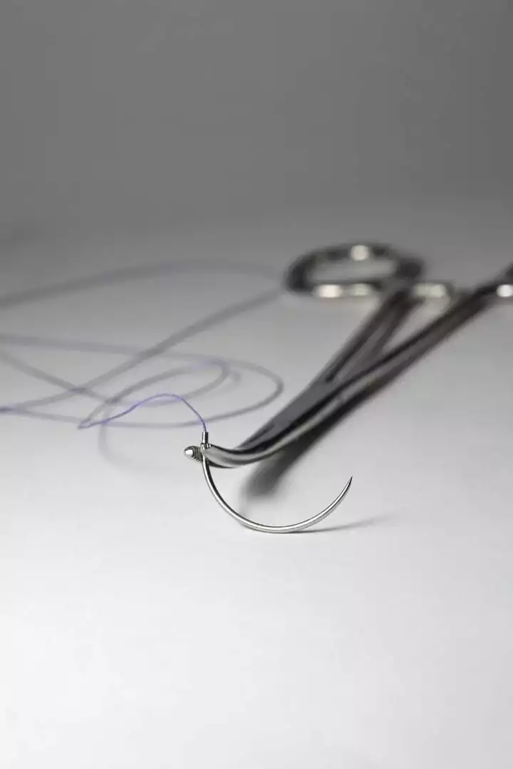
- Swaged needles, with the suture permanently attached, are generally preferred over eyed needles.
- Curved needles are the most used; 3/8 and 1/2 circle are the most common curves.
- Taper (round-bodied) points suit delicate tissue; reverse cutting needles suit skin and other tough tissue. For the gut, Guelph recommends a taper or taper-cut needle.
On suture material (WCVM, classification of sutures): absorbable sutures lose most of their strength within 60 days and are broken down by the body. Nonabsorbable sutures keep their strength longer and can stay buried, but should not sit in the lumen of the bladder or biliary system, where they can seed stones. Monofilament has minimal tissue drag but needs extra throws for a secure knot; braided multifilament holds knots better but can harbor bacteria between its fibers.
For the stomach, intestine and bladder, Guelph recommends monofilament absorbable suture and rules out chromic gut, which loses strength very quickly in the gut and in urine.
Other suture patterns you will meet
Several patterns can suit the same incision; species, patient and surgeon preference all shape the choice. The rest of the set from the same surgery labs:
Interrupted patterns
- Simple interrupted: each stitch is a separate bite across the incision, tied and cut. Losing one or two stitches does not usually open the closure, but it takes longer. It suits normal tension, not significant tension.
- Buried knot: the needle goes in deep and out superficially on one side, then in superficially and out deep on the other, so the knot sits down in the tissue. Guelph uses it to start and end subcutaneous lines.
- Cruciate: two simple interrupted bites crossed into an X and tied once. WCVM counts it as a tension suture.
- Horizontal mattress: an appositional pattern for normal tension. WCVM warns its effect on the blood supply is major if it is overtightened.
- Vertical mattress: an everting tension suture, with a wide outer bite and a narrow inner bite.
Continuous patterns
- Simple continuous: like simple interrupted, but the suture is not cut between bites, so there is a knot only at each end.
- Ford interlocking: a simple continuous in which the needle passes through the previous loop before each new bite.
- Intradermal: bites run parallel to the incision inside the dermis, with buried knots at both ends.
- Purse-string: Cushing-type bites around a stump or tube, pulled tight.
- Lembert and Utrecht: two more inverting patterns for hollow organs. The UMN notes list both alongside the Cushing.
- Skin staples: not a suture pattern, but an alternative for skin. After a C-section, Guelph prefers staples or an intradermal closure because the puppies will be nursing.
For the full textbook treatment of suture patterns and soft tissue surgery, our list of the best veterinary books and study material is a good place to start.
Sources
- University of Guelph, Veterinary Surgery Online: Cushing Pattern
- University of Guelph, Veterinary Surgery Online: Gastrotomy Closure
- University of Guelph, Veterinary Surgery Online: Cystotomy Closure
- University of Guelph, Veterinary Surgery Online: Close the Uterus in 2 Layers
- University of Guelph, Veterinary Surgery Online: Intestinal Apposition and Closure
- University of Guelph, Veterinary Surgery Online: Suture Materials
- University of Guelph, Veterinary Surgery Online: Simple Interrupted, Simple Continuous, Ford Interlocking, Intradermal, Purse-String, Cruciate, Burying a Knot
- Western College of Veterinary Medicine, University of Saskatchewan: Continuous Patterns
- WCVM: Suture Patterns
- WCVM: Interrupted Suture Patterns
- WCVM: Needles
- WCVM: Classification of Sutures
- University of Minnesota, Large Animal Surgery: Inverting Suture Patterns
- Lundberg RK et al.: The Connell Stitch, a two-generation contribution to gastrointestinal surgery, The American Surgeon (2023)
- Hildreth BE et al.: Single-layer continuous Cushing vs simple continuous cystotomy closure in rats, American Journal of Veterinary Research (2006)
- Ellison GW et al.: Intestinal surgery in small animals, Veterinary Surgery (2019)
- Lee WL et al.: One-layer vs two-layer (simple continuous/Cushing) jejunoileal anastomosis in horses, Veterinary Surgery (2012)
- Singh H et al.: Colonic anastomosis in calves, Acta Veterinaria Hungarica (1989)
This article is for general information and is not a substitute for advice from your veterinarian.