
Dermatology terms describe what a skin change looks like before anyone names the disease behind it. Primary lesions come straight from the disease (a macule, papule, pustule or wheal), while secondary lesions develop later, as the lesion ages or the pet scratches and licks (an epidermal collarette, crust, erosion or lichenified patch). Learn the words below and you can read, and write, the skin section of almost any exam note.
Veterinary dermatology is a specialty in its own right. The skin is the largest organ of a dog's body and makes up roughly 12 to 24% of body weight, depending on species and age. The lesion definitions here follow veterinary teaching sources, mainly the Royal Veterinary College's Diagnosis in Dermatology teaching module and the Merck Veterinary Manual. If your course notes use a slightly different list, go with your course for the exam.
Primary vs Secondary Skin Lesions at a Glance
Group | Lesions | What it tells you |
|---|---|---|
Primary | Macule, patch, papule, plaque, nodule, tumor, vesicle, bulla, pustule, wheal | The disease itself made this. Most useful for building the differential list |
Secondary | Epidermal collarette, erosion, ulcer, excoriation, fissure, lichenification, hyperkeratosis, scar, callus | It came later: from an older primary lesion, from healing, or from the pet's own scratching, licking and rubbing |
Can be either | Scale, crust, alopecia, comedo, follicular cast, hyperpigmentation, hypopigmentation | Depends on the disease. Read it together with the rest of the picture |

The split is a teaching tool, not a law. The same lesion can be primary in one disease and secondary in another. Alopecia is a good example: in a hypothyroid dog the hair loss comes directly from the hormone problem (primary), while in a cat with flea allergy the cat pulls the hair out itself (secondary).
One more trap for students: a "secondary lesion" is not the same thing as "secondary pyoderma." Secondary pyoderma means a bacterial infection on top of another skin disease, such as an allergy or demodicosis, and it often shows up with primary lesions like papules and pustules.
Primary Skin Lesions
Most primary lesions are sorted by two questions: is it flat or raised, and what is inside it? Size then picks the name.
Type | Smaller than 1 cm | Larger than 1 cm |
|---|---|---|
Flat color change | Macule | Patch |
Solid and raised | Papule | Plaque (flat-topped) or nodule (deeper) |
Raised with clear fluid | Vesicle | Bulla |
Raised with pus | Pustule | Pustule (no size split) |
Raised with dermal fluid (edema) | Wheal | Wheal |
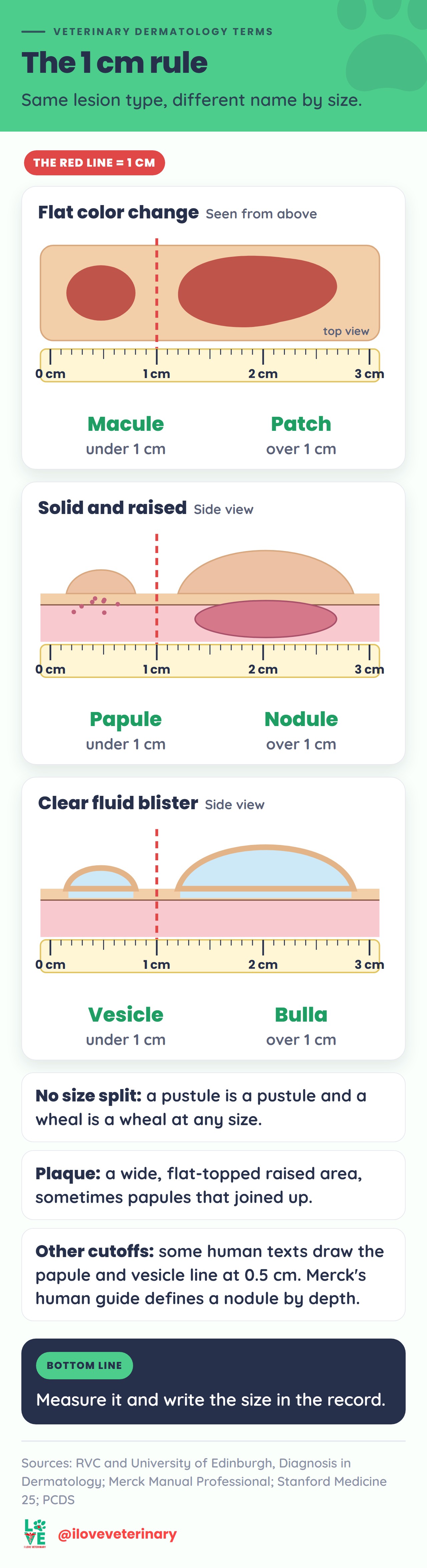
- Macule: a flat, circumscribed change in skin color smaller than 1 cm. The color can come from extra or lost pigment, redness (erythema) or bleeding. Red macules are common in allergic skin disease.
- Patch: a macule larger than 1 cm. Pinpoint macules caused by bleeding are petechiae; bleeding patches over 1 cm are ecchymoses.
- Papule: a small, solid bump smaller than 1 cm, usually made of inflammatory cells. Flea bite reactions in dogs and cats and canine pyoderma both commonly cause papules.
- Plaque: a larger, flatter raised area, sometimes formed when papules join together. The eosinophilic plaque in cats with allergic skin disease is the classic example.
- Nodule: a solid bump larger than 1 cm that usually reaches into the deeper layers of the skin. Causes include tumors and collections of inflammatory cells, so a nodule always needs a closer look by the vet.
- Tumor: a large mass involving the skin. The word is often used for cancer, but technically an inflammatory mass can be a tumor too.
- Vesicle: a small blister filled with clear fluid, smaller than 1 cm. Vesicles are rare in animals and usually turn into erosions and ulcers, so you are more likely to see what they leave behind.
- Bulla: a blister larger than 1 cm. Causes of blisters include viral infections, autoimmune disease and irritant chemicals.
- Pustule: a small raised spot in the epidermis filled with pus. In dogs, most pustules come from staphylococcal infection (pyoderma). Sterile pustules point to rarer immune-mediated disease, most importantly pemphigus foliaceus in dogs, cats and horses.
- Wheal: a raised, well-defined swelling caused by fluid in the dermis, usually from mast cells releasing their contents. The condition is called urticaria (hives). Insect bites, stings, drug reactions and allergies can all cause them, and they are seen most often in horses.
Why Size Cutoffs Differ Between Textbooks
The RVC veterinary module uses 1 cm to split macule from patch, papule from nodule and vesicle from bulla, and Stanford's human dermatology guide does the same. The Merck Manual's professional (human) guide puts the macule, papule and vesicle limit at about 10 mm too, but it defines a nodule by depth, as a lesion reaching into the dermis or the tissue under the skin, rather than by size. Some human references, such as the UK Primary Care Dermatology Society, draw the papule/nodule and vesicle/bulla lines at 0.5 cm. Learn the number your course uses, and in a record, write down the measurement: a lesion's size in centimeters tells the next reader more than its name.
Lesions That Can Be Primary or Secondary
- Scale: loose flakes of the skin's outermost, keratin-filled layer. Scaling can be a primary keratinization problem such as idiopathic seborrhea, or follow mites (cheyletiellosis), infection, ringworm or allergy.
- Crust: dried exudate on the skin surface: serum, blood, pus or a mix. Crusts are common in pustular diseases such as pyoderma and pemphigus foliaceus and over erosions and ulcers. A crust is not a scale, even if both look "flaky."
- Alopecia: partial or complete hair loss. Describe the pattern too: focal, multifocal, regional, diffuse or generalized. Our guide to alopecia in dogs covers the causes.
- Comedo (plural comedones): a hair follicle stretched open and plugged with keratin, the animal version of a blackhead. Comedones are most often seen in dogs, for example with demodicosis, and also appear in feline acne.
- Follicular cast: a collar of keratin debris around the hair shaft where it leaves the follicle. It points to a problem with how the follicle makes keratin and often appears with comedones, for example in sebaceous adenitis.
- Hyperpigmentation / hypopigmentation: skin that is darker or paler than normal. Dark skin often follows chronic inflammation or rubbing.
Secondary Skin Lesions
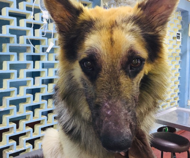
- Epidermal collarette: a ring of peeling scale with a clear edge, usually left behind when a pustule, papule or vesicle breaks. Collarettes are a hallmark of superficial pyoderma in dogs and can darken over time.
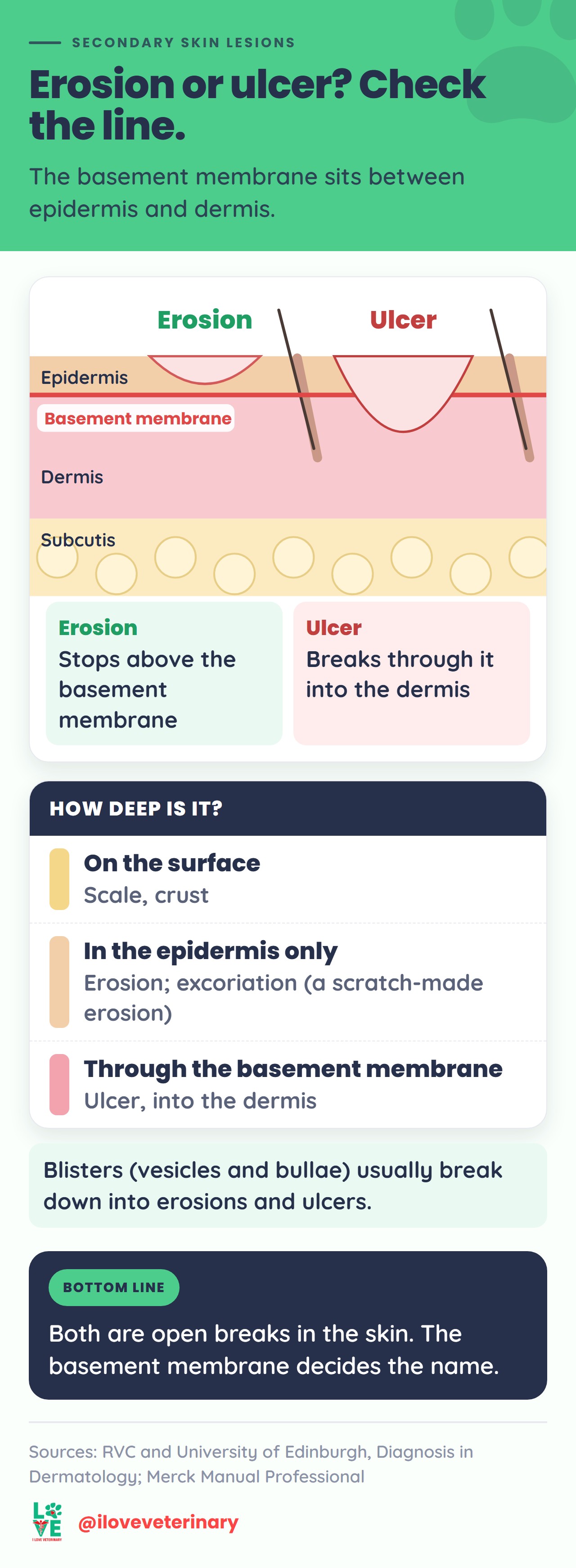
- Erosion: a shallow break in the epidermis that does not go through the basement membrane (the boundary between the epidermis and the dermis).
- Ulcer: a deeper break that goes through the basement membrane into the dermis. Blisters usually turn into erosions and ulcers as they break down.
- Excoriation: a scratch-type erosion, often linear, caused by the pet scratching or rubbing. It is a sign of itch.
- Fissure: a crack or split in the skin.
- Lichenification: thickened, leathery skin with exaggerated skin markings, caused by chronic rubbing or inflammation. The name comes from tree lichen. It is a typical sign of long-term itch, for example in dogs with atopic dermatitis or flea allergy.
- Hyperkeratosis: a thickened stratum corneum (the dead, horny top layer of the epidermis), seen for example on footpads and ear margins.
- Scar: fibrous tissue that replaces normal skin after injury. Deep burns, for example, heal with hairless scars.
- Callus: thickened, hairless skin over a bony pressure point. The classic spot is the elbow of a large or giant breed dog that lies on hard floors.
Distribution and Pattern Words
Where the lesions sit, and the shape they make, narrows the list of causes as much as the lesion type does.
- Localized, multifocal, generalized: one area, several separate areas, or most of the body.
- Symmetrical: both sides affected about equally, which often suggests a systemic (body-wide) cause.
- Annular: ring-shaped, which usually means the disease is spreading outward, as in pyoderma or ringworm.
- Linear: in a line, pointing to outside trauma, a blood vessel or a nerve path.
A few classic patterns students are expected to know:
Pattern | Think about |
|---|---|
Pustules on the belly and inner thighs of a dog | Superficial pyoderma |
Pustules on the face and ear flaps | Pemphigus foliaceus |
Lesions mostly on the back half of the body | Flea allergy dermatitis |
Itchy paws, face, ears, armpits and belly | Atopic dermatitis |
More Veterinary Dermatology Terms (A to Z)
- Abscess: a walled-off pocket of pus in the tissue, often just under the skin. In cats, small, deep bite wounds often become infected and are treated as abscesses.
- Atopy (atopic dermatitis): in dogs, a hereditary, typically itchy inflammatory skin disease driven mainly by T cells, in which a weak skin barrier, sensitization to allergens and an imbalance of skin microbes all play a part. That is the 2023 definition from the International Committee on Allergic Diseases of Animals. More in our atopic dermatitis in dogs guide.
- Cyst: a cavity lined by epithelium, filled with fluid or solid material. Many lumps get called cysts that are not, which is why a vet checks every new lump.
- Dermatitis: inflammation of the skin.
- Epidermis vs epithelium: the epidermis is the outer layer of the skin. Epithelium is the type of tissue that covers body surfaces; the epidermis is one kind (a stratified squamous, keratinizing epithelium).
- Otitis externa: inflammation of the external ear canal, outside the eardrum. The ear flap may or may not be involved. It is one of the most common reasons dogs and cats see a vet.
- Pododermatitis: inflammation of the feet or paws. The pads, the nail beds and the skin on the top and underside of the paw can all be involved.
- Pruritus: itch. (Spelled pruritus, not "pruritis.") Pruritic means itchy.
- Pyoderma: literally "pus in the skin," a bacterial skin infection. In dogs the main culprit is Staphylococcus pseudintermedius, and the infection is usually secondary to something else, such as parasites, allergies or trauma.
- Seborrhea: a disorder of keratinization that shows as scaling, greasy skin or both. Primary seborrhea is a rare inherited disease; secondary seborrhea, caused by another problem such as allergies or hormonal disease, is far more common.
- Wood's lamp: an ultraviolet lamp. Hairs infected with Microsporum canis, a common cause of ringworm in cats, can glow apple green under it, which helps the vet choose hairs to test.

From Lesion to Diagnosis
Describing lesions is not busywork. The type and distribution of lesions decide which diseases go on the differential list, and that decides which tests come next. Common next steps include examining skin scrapings and hairs under the microscope, cultures of hair or skin swabs, skin tests, blood and urine tests, and biopsies. Many skin diseases look alike, so getting to a diagnosis often takes more than one visit.
When a case is stubborn, the general practice vet may refer to a board-certified veterinary dermatologist. In the US those specialists are Diplomates of the American College of Veterinary Dermatology, an AVMA-recognized specialty organization.
To see these lesions in real cases, our 10 common dog skin problems (with pictures) matches each sign to its likely causes. And for the shorthand that surrounds the skin section of a chart (Hx, PE, R/O, Tx), keep our veterinary abbreviations cheat sheet open next to this one.
Quick Questions
What is the difference between a macule and a patch?
Only size. Both are flat color changes you can see but not feel; a macule is smaller than 1 cm and a patch is larger.
Is a crust a primary or a secondary lesion?
It can be either. A crust forms when exudate dries, so it often follows a broken pustule or an erosion, but in some diseases crusting is part of the main picture from the start.
What is an epidermal collarette?
A ring of peeling scale left where a pustule, papule or vesicle has broken. In dogs it is one of the most typical signs of superficial pyoderma.
What is the difference between an erosion and an ulcer?
Depth. An erosion stays in the epidermis, above the basement membrane; an ulcer goes through it into the dermis.
Sources
- RVC and University of Edinburgh: Diagnosis in Dermatology, Skin Lesions module
- Animal Health Australia: Gross morphology of lesions (veterinary teaching slides)
- Merck Veterinary Manual: Atopic Dermatitis in Dogs
- Merck Veterinary Manual: Pyoderma in Dogs and Cats
- Merck Veterinary Manual: Seborrhea in Animals
- Merck Veterinary Manual: Otitis Externa in Animals
- Merck Veterinary Manual: Dermatophytosis in Dogs and Cats
- Merck Veterinary Manual: Hygroma in Dogs
- Merck Veterinary Manual: Management of Specific Wounds in Small Animals
- Merck Veterinary Manual: Structure of the Skin in Dogs
- Merck Veterinary Manual: Diagnosis of Skin Disorders in Dogs
- VCA Animal Hospitals: Pododermatitis in Dogs
- Merck Manual Professional Version: Description of Skin Lesions
- American College of Veterinary Dermatology
- Stanford Medicine 25: The Dermatology Exam, Learning the Language
- Primary Care Dermatology Society: Dermatology Dictionary
This article is for general information and is not a substitute for advice from your veterinarian.