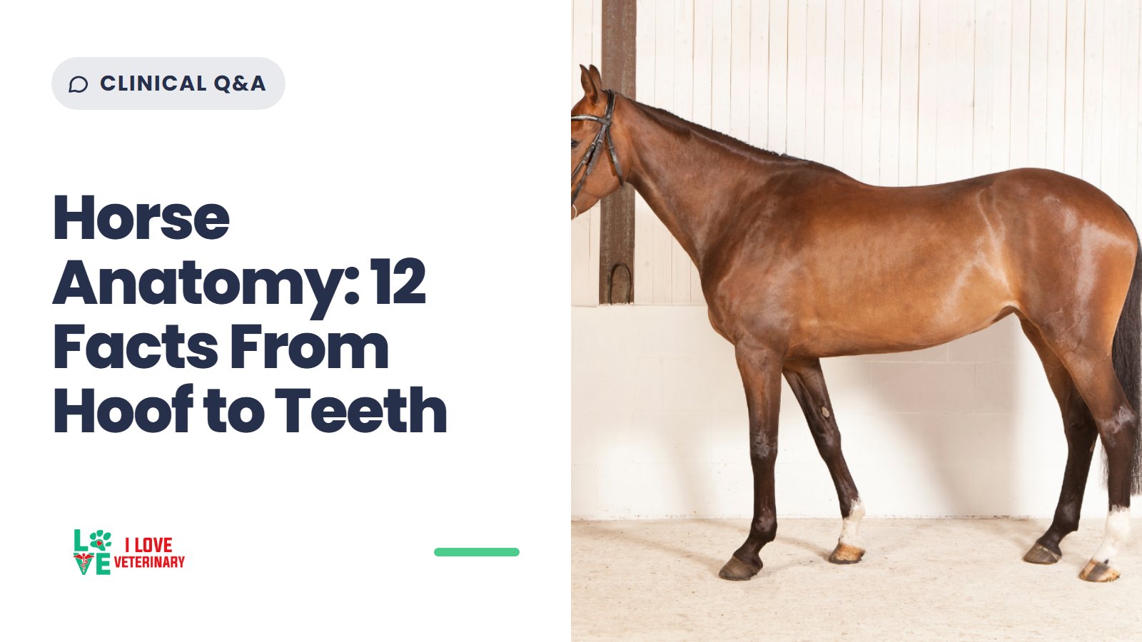
Horses are large prey animals built for two jobs: grazing for most of the day and running when something startles them. You can see both in their bodies, from a gut designed for a steady flow of forage to legs that end in a single toe.
Horses have been evolving for millions of years, but the domestic horse is a much more recent story. A 2021 genome study in Nature traced modern domestic horses to the lower Volga-Don region of the western Eurasian steppes and found that they spread rapidly across Eurasia from about 2000 BC. Horses were once used for transport and even battle; today most are kept for work, sport, show and companionship.
This article is an anatomy reference, not a care guide. For questions about a specific horse's health, your equine veterinarian is the right person to ask.

The Skeleton and Legs
A horse's skeleton has about 205 bones. The exact number varies a little by breed, and Arabian horses can have fewer bones than other breeds.
The legs are where horse anatomy gets strange. Each leg ends in a single digit, so a horse is standing on the equivalent of one finger. The coffin bone inside the hoof is the distal phalanx, the same bone that sits in the tip of your finger. In the foreleg, the radius and ulna are fused together, so a horse cannot rotate its forearm the way a person can.

Horses can also rest on their feet thanks to the stay apparatus, a system of tendons and ligaments that keeps the limbs straight with very little muscle work. In the hind leg, the kneecap (patella) hooks over the end of the thigh bone (femur) and locks the stifle joint. The lock is not completely passive, though. A 2003 study in the Journal of Anatomy found that one thigh muscle, the vastus medialis, stays active while the leg bears weight in the locked position, at an estimated tension of less than 2% of the force the leg would need without the lock.
The Hoof
The hoof wall grows down from the coronary band, the ring of tissue where the hairline meets the top of the hoof, at about 6-9 mm (1/4 to 1/2 inch) a month. At that pace a horse grows a new hoof in about a year. It also means damage to the wall cannot be repaired from above: it has to grow out.
Inside and under the wall sit several structures with distinct jobs:
- Frog: the wedge-shaped, rubbery pad on the underside of the hoof. It is about 50% moisture, absorbs shock, and helps with heel expansion and blood circulation.
- Digital cushion: a tough, elastic pad above the frog, visible from the outside as the bulbs of the heels.
- Lateral cartilages: cartilage plates on either side of the foot.
- Laminae: the layers of tissue that attach the coffin bone to the inside of the hoof wall.
- White line: the junction of the wall and the sole.
- Navicular bone: a small, shuttle-shaped bone next to the coffin bone, closer to the heel.
For more horse vocabulary, from withers to pastern, see our guide to common horse terms.
The frog is often described as a pump that sends blood back into the leg. The fuller picture involves three structures working together: each time the foot lands, the frog and digital cushion compress and squeeze veins against the lateral cartilages, which pushes blood back up the leg toward the heart.


The laminae explain why hoof problems can turn serious fast. Laminitis is the inflammation and then separation of these laminae. When they fail, the coffin bone can rotate or sink inside the hoof. Merck describes acute laminitis as an emergency that can become life-threatening, so a horse with suspected laminitis needs a veterinarian promptly.
Teeth and Aging
Mature stallions have 40 to 44 teeth and mature mares have 36 to 40. The difference is mostly the canine teeth, which are often absent in mares. Foals have 24 deciduous (baby) teeth, and all permanent teeth are usually in place by about 5 years of age.
A horse's age can be estimated from which teeth have erupted and how the lower incisors have worn. It is an estimate, not an exact measure, so a dental age is a useful clue rather than a birth certificate.
Organ Functions in Horses' Anatomy
A horse's digestive tract is built for small amounts of forage eaten almost all day, and that design shapes much of the organ list below.
- Trachea: the passage carrying air to and from the lungs.
- Heart: pumps blood throughout the body. It sits near the horse's left elbow.
- Lungs: bring oxygen into the blood.
- Diaphragm: separates the thoracic (chest) and abdominal cavities.
- Stomach: small for an animal this size, holding about 2 to 2.5 gallons (8 to 10 liters). Food moves through it quickly, which suits continuous grazing.
- Small intestine: with the stomach, this is the foregut, where enzymes do the digesting. One section of it, the jejunum, is about 65 feet (19.5 meters) long.
- Cecum: a large fermentation vat that can hold 7 to 8 gallons (27 to 30 liters) of food and water. Bacteria here start breaking down fiber.
- Colon: the large and small colon continue the work of the hindgut, where bacteria ferment fiber into volatile fatty acids, and form fecal balls.
- Bladder: stores urine.
The intestines also bend and change diameter along the way, and those turns and narrow points raise the risk of impactions.

Why Horses Can't Vomit
Where the esophagus meets the stomach, a horse has a functional one-way valve. Gas and fluid can move into the stomach but not back out, so horses can neither regurgitate nor vomit. If the stomach fills with gas or fluid, it can rupture. According to the Merck Veterinary Manual's overview of colic, a veterinarian passing a stomach tube to relieve that pressure may save the horse's life.
That is one reason to call your veterinarian right away about colic, the general term for abdominal pain in horses, rather than wait and see.
Airway and Breathing
In a horse, the soft palate (the soft back part of the roof of the mouth) forms a seal around the epiglottis, the flap that guards the entrance to the windpipe. The seal opens only when the horse swallows. As a result, horses are obligate nasal breathers: under normal conditions they cannot breathe through their mouths, and they cannot pant.
Deeper in the respiratory tract, two conditions are worth knowing by name. Equine asthma is a chronic, non-infectious disease of adult horses involving inflammation of the airways, and respirable dust in hay or bedding is a known trigger. Exercise-induced pulmonary hemorrhage (EIPH) is bleeding into the lungs after strenuous exercise, caused by tiny lung capillaries rupturing. It occurs in most racehorses.
A veterinarian diagnoses and manages both.
Eyes and Vision

Horses have among the largest eyes of any land mammal. The eyes sit on the sides of the head, which gives a horse near-complete peripheral vision. The only blind spots are created by the horse's own body: one behind it and one directly in front of the forehead. Straight ahead, the fields of view from both eyes overlap by about 65 to 80 degrees, which gives the horse its binocular vision.

Interesting Facts About Horse Anatomy
- Horses have among the largest eyes of any land mammal.
- A horse's skeleton has about 205 bones, and the count varies slightly by breed.
- Horses cannot vomit. A one-way valve where the esophagus meets the stomach lets gas and fluid in but not out.
- Horses make saliva only while they chew. The parotid glands alone produce an estimated 12 liters (about 3 gallons) a day in an average horse.
- When a horse curls back its upper lip (the flehmen response), it is moving scent molecules, including pheromones, into the vomeronasal organ, a specialized scent organ once known as Jacobson's organ. Stallions do it most, but mares and geldings do it too.
- A horse can breathe only through its nose under normal conditions, because the soft palate seals off the mouth.
- The average gestation period for a mare is about 340 days, roughly 11 months.
- Horses don't have seven "blood types." They have seven recognized blood group systems (A, C, D, K, P, Q and U) with more than 30 red blood cell antigens between them.
- The frog does not pump blood on its own. It works with the digital cushion and lateral cartilages to push blood back up the leg.
- A horse grows a whole new hoof wall in about a year, so any damage has to grow out from the coronary band.
- A horse sees almost all the way around itself. Its blind spots are directly behind it and right in front of its forehead.
- Mature stallions have 40 to 44 teeth and mares 36 to 40, and age estimates from the lower incisors are just that: estimates.
If horse anatomy is the part of veterinary medicine that grabs you, read our guide on how to become an equine veterinarian.
Originally written by Amber LaRock. Updated by the I Love Veterinary editorial team.
Sources
- Merck Veterinary Manual: Overview of Colic in Horses
- Merck Veterinary Manual: Introduction to Digestive Disorders of Horses
- Merck Veterinary Manual: Dental Development of Horses
- Merck Veterinary Manual: Laminitis in Horses
- Merck Veterinary Manual: Asthma in Horses
- Merck Veterinary Manual: Exercise-Induced Pulmonary Hemorrhage in Horses
- Merck Veterinary Manual: Management of Reproduction in Horses
- Frontiers in Veterinary Science: Horse vision through two lenses: Tinbergen's Four Questions and the Five Domains
- NC State College of Veterinary Medicine: Ophthalmology
- Kentucky Equine Research: The Horse's Mouth: Understanding the Soft Palate
- Kentucky Equine Research: Saliva Production and Function in the Horse
- Kentucky Equine Research: Flehmen: The Horse with the Upturned Lip
- Kentucky Equine Research: The Horse's Hoof
- Journal of Veterinary Internal Medicine: Comparison of Tube, Gel, and Immunochromatographic Strip Methods for Evaluation of Blood Transfusion Compatibility in Horses
- UC Davis Center for Equine Health: Parts of the Horse
- Rutgers Equine Science Center: Not So Scary Skeletons
- Journal of Anatomy: The equine hind limb is actively stabilized during standing
- Nature: The origins and spread of domestic horses from the Western Eurasian steppes
This article is for general information and is not a substitute for advice from your veterinarian.