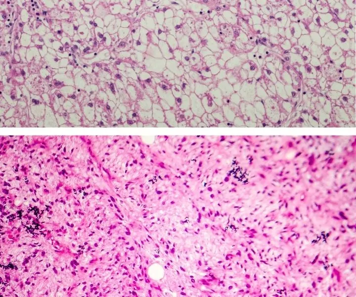
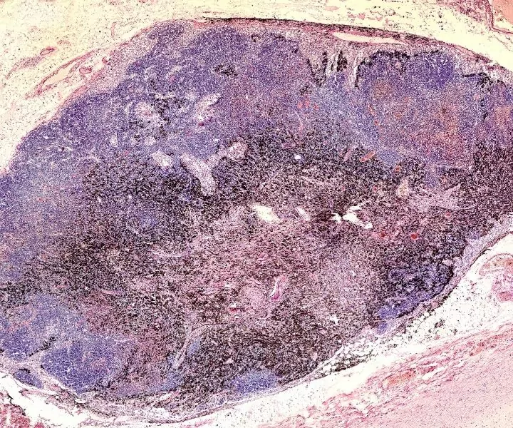
Carcinoma vs Sarcoma -What Are They?
When discussing carcinoma vs sarcoma in dogs, this implies cancers that develop when individual cells that would usually die and be replaced instead grow out of control and form new, abnormal cells.

The body is ordinarily able to stop this process when it occurs, but occasionally this control mechanism doesn’t function, allowing for uncontrolled development. These extra cells may form a mass called a tumor, although some cancers, such as leukemias, do not.
There are many different types of tumors. Today we are going to focus on carcinomas vs. sarcomas in dogs. Although these tumors are different, they share the same part of the word, -oma. This means tumor, and there are many types, including adenoma, carcinoma, sarcoma, and lymphoma.
A carcinoma (carcino- means cancer) develops from the uncontrolled growth of skin or tissue cells that line the body’s internal organs, e.g., liver or kidney.
A sarcoma (sarco- means flesh or muscle) develops from the uncontrolled growth of cells in the body’s connective tissue, e.g., fat, nerves, bones, and muscles.
These two types of cancer are very different and have varied prognoses depending on the kind, which is discussed in more detail below.
Types of Carcinomas in Dogs
There are no benign tumors that are carcinomas; by definition, carcinomas are malignant tumors. In general, carcinomas usually do not spread to other organs but instead are locally invasive.
Carcinomas are made from cells that line organs. This means they can develop in many locations such as the skin, nasal cavity, chest cavity, and bladder.
Tumors that are external or easily felt through the skin can be sampled in conscious animals to help aid in diagnosis. A fine needle aspirate is performed, whereby a small needle is passed into the tumor, and negative pressure is applied to the attached syringe. The needle is redirected a few times to suck up cells from the mass. These cells are then released onto a glass slide and sent to the lab for analysis.

A pathologist will perform cytology whereby they look at the cells collected under a microscope and determine the type of tumor that the dog has. This diagnosis can help a veterinarian decide on the next step in diagnosis or treatment in the canine.
Unfortunately, cytology isn’t always diagnostic, and a biopsy may be required. Also, if a mass is internal and cannot be accessed through the skin, the animal will likely need to be anesthetized and the skin cut open to expose the mass.
When the dog is being operated on for a diagnosis to be made, a biopsy is often much more helpful than a fine needle aspirate as the chance of diagnosis is higher, and repeated surgeries for diagnosis would most likely be avoided.
Examples of Carcinomas in Dogs
Squamous cell carcinoma
A tumor of the skin. Typically, squamous cell carcinomas appear as a single lesion in one location by itself. They often develop on skin with exposure to a lot of sunlight, such as pale pink skin on the ears of white dogs.
Boxer dogs, Poodles, and Dalmations are at increased risk of developing squamous cell carcinomas. The prognosis varies depending on the type, if it can be removed and how much it has invaded into local structures of the body.

Transitional cell carcinoma
This is a tumor of the lining of the bladder and urethra. It is most commonly found in senior small breed dogs such as Dachshunds and West Highland White Terriers. It is ultimately fatal as it continues to grow, and it can block the urethra or ureter, preventing urine flow and causing an obstruction.
Hepatocellular carcinoma
This type of carcinoma is a tumor of the liver. It is the most common primary liver tumor in dogs. They are most commonly diagnosed through palpation of the liver on physical examination by veterinarians. Hepatocellular carcinomas have not been linked to specific breeds, and the prognosis varies on whether the tumor can be removed or not.
Mammary adenocarcinoma
These are a type of tumor formed in the mammary tissue. There are two types, tubular and papillary. These types of mammary tumors have a poor prognosis and are most common in unneutered dogs.
Although these types of mammary tumors are malignant, 50% of mammary tumors in dogs are actually benign.
Types of Sarcomas in Dogs
Sarcomas can be malignant or not; the most common group of sarcomas we see in dogs are soft tissue sarcomas.
A diagnosis from cytology can tell you what sort of sarcoma your dog has, but it often requires histology from a biopsy to determine the grade. A biopsy involves making an incision into the tumor and either removing a small piece (incisional biopsy) or the whole mass (excisional biopsy) and sending it, preserved in formalin, to the lab for a pathologist to examine under a microscope.
Histology grades vary from I to III, and the grade can help us know the prognosis and treatment required for a specific tumor.
To further learn about a dog’s tumor, the sarcoma might be staged, and this involves determining how far the cancer has spread through the body and how big it is. To stage a tumor, it is often necessary to perform cytology on nearby lymph nodes, as this is where tumors typically spread first.
If cancer cells are seen in the lymph nodes, this is classified as a high stage and is the worse possible prognosis. Imaging can also be used to see if the tumor has spread to other organs. For example, osteosarcomas will often spread to the lungs; seeing metastatic masses on x-ray will mean that the tumor is at a higher stage.

The size of a tumor will also determine its stage. A larger tumor will have a higher number. Soft tissue sarcomas can be staged from I to IV.
Examples of Sarcomas in Dogs
Liposarcomas
These develop from fat cells in older dogs. Liposarcomas are uncommon and malignant. They are often firm to the touch and have an ill-defined border. They are locally invasive and rarely spread to other organs. The average survival time is six to12 months and is not associated with any specific breed.
Osteosarcomas
These are primary bone tumors of the limbs and occasionally develop in the skull, spine, or rib cage. They are the most common primary bone tumor in dogs and are most prevalent in large- or giant-breed dogs such as Great Danes, Irish Wolfhounds, and Rottweilers. Osteosarcomas have a poor prognosis as they commonly spread to the lungs.
Hemangiosarcoma
This is a type of tumor that develops from cells that line the blood vessels. This means it can show up in various organs, including the heart and liver. Hemangiosarcomas are relatively common and makeup between 5-7% of all tumors in dogs.
It is most predisposed in middle-aged dogs. The breeds that are commonly affected include German Shepherds, Golden Retrievers, and Boxers.
Neurofibrosarcomas
They are a malignant type of tumor that develops on the peripheral nerve sheaths, that is, the protective lining that covers nerves in the body.
Neurofibrosarcomas are most commonly on the nerve sheaths that surround the bundle of nerves that innervate the front and back legs. They are slow-growing and invade locally but are unlikely to spread to other organs.
Conclusion
As you can see in this article, Carcinomas and Sarcomas are both tumors but differ in many different ways. Carcinomas are more aggressive tumors that are always malignant and develop from cells that line organs. Carcinomas are locally invasive.
Sarcomas can be malignant or benign. They develop from connective tissue cells and can spread throughout the body. It is essential that you get your dog examined by a veterinarian if you ever notice an abnormal growth or changes in your animal’s behavior.
If the veterinarian is concerned about the possibility of tumors, tests can be performed to aid in diagnosis. Both types of tumors can be treated in different ways, including surgery, chemotherapy, and radiation.
