What is Elbow Dysplasia in Dogs?
Elbow dysplasia in dogs is the abnormal development of the elbow joint. The condition affects mainly young, large breed dogs because of their rapid growth rates. The bone growth, cartilage formation, and joint conformation all become adversely affected, and the elbow joint becomes dysfunctional.
Dogs can begin to show signs of lameness, pain, and gait abnormalities from four to eight months old. The dog’s elbow joint is complex, and the condition can involve multiple developmental abnormalities.

Where is a Dog’s Elbow?
The elbow joint comprises three bones, the radius, the ulna, and the humerus. The joint’s congruency (the precise fit) is essential for the elbow to function normally.
The Elbow – Where Does it All Go Wrong
Elbow dysplasia (ED) is a collection of several conditions that affect the normal function of the joint in different ways. One or several conditions can affect the joint and cause canine elbow dysplasia.
The exact causes of ED are unclear, but multiple factors, including genetics, nutrition, trauma, and cartilage development, seem to be interlinked in the condition.
Fragmented Medial Coronoid Process (FCP)
The coronoid process is a small bony protrusion at the end of the ulna. FCP in dogs occurs when the medial process does not form correctly. The abnormality leads to crack formations and separation from the rest of the ulna.
Osteochondritis Dissecans (OCD)
The medial humeral condyle undergoes inflammatory changes resulting in the cartilage below the bone dying. The cartilage dies because of a lack of blood flow. The brittle dead cartilage sometimes breaks off and causes joint mice that hinder joint mobility and cause pain.
Ununited Anconeal Process (UAP).
The anconeal process has a growth plate of cartilage that develops separately from the ulna. The plate closes as the dog reaches four months of age, and the two bones become fused. Abnormal development of the elbow can lead to pressure on the anconeal process, which affects its fusion to the ulna.
If the anconeal process fails to fuse to the ulna, the joint becomes afflicted with an ununited anconeal process. A UAP leads to joint incongruity and results in the development of arthritis.
Medial Compartment Disease
With elbow dysplasia, the dog’s elbow develops abnormally. It places too much pressure on one side of the joint and results in cartilage loss. Medial compartment disease results from the loss of cartilage on the medial coronoid process and the condyle of the humerus.
Cartilage loss and inflammation result in arthritis and permanent joint damage. These changes lead to decreased joint mobility, pain, and possibly even loss of joint function. This form of elbow dysplasia carries a very poor prognosis.
Signs of Elbow Dysplasia in Dogs
The initial signs of elbow dysplasia can be subtle. Still, it is essential to diagnose the condition early to ensure that the joint does not undergo any permanent changes. Dogs with ED can show low-grade, intermittent lameness.
Owners need to monitor their pets closely for signs of ED, especially if their pets are a high-risk breed or have parents with ED.
Dogs can start with symptoms of ED at an early age. The average onset of clinical signs varies from 5 months to 5 years. Afflicted animals have a gradually progressive lameness in the forequarters that may worsen after exercise.
The following symptoms could indicate an underlying issue:
- Occasional limping that becomes exaggerated after exercise or when first standing up.
- Abnormal conformation where one or both paws rotate inwards and the elbows rotate outwards.
- The elbow is often stiff, or the dog struggles to achieve the full range of motion of the joint.
- An abnormal gait when walking in a straight line.
- Decreased or under-developed muscle tone around the shoulder.
- Crepitus or clicking of the elbow when moved.
- Reluctance to exercise or play.
How is Elbow Dysplasia in Dogs Diagnosed?
If your pet suffers from forelimb lameness, a vet will first start with a complete clinical history. This includes breeding, nutrition, history of any previous trauma, and exercise habits. The vet will then perform a thorough clinical exam and determine if there is a need for further diagnostic tests.
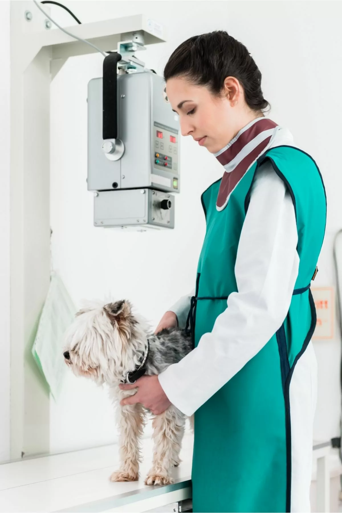
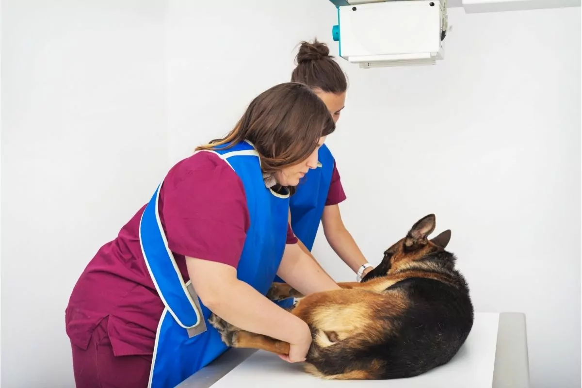
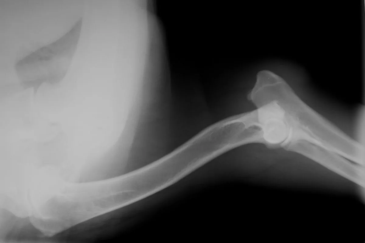
The diagnosis of ED requires a radiological evaluation of the elbow joint. CT and MRI scans are more accurate, but they are also more expensive. A basic assessment of the elbow can reveal osteoarthritic changes, joint mice (small fragments of cartilage or bone), or an ununited anconeal process.
Radiographs can be taken at a GP vet practice and sent to a veterinary radiologist if the owner requires an ED certificate. ED- screening is recommended at 24 months for large breeds dogs, but a vet can assess a dog at around 18 months.
The assessment of canine elbow dysplasia radiographs is according to a grading system as per the table below.
| Elbow Dysplasia Scoring | Radiographic findings | |
| 0 | Normal elbow joint | Normal elbow joint. No evidence of incongruency, sclerosis, or arthrosis. |
| 1 | Mild arthrosis | Presence of osteophytes < 0.08 inches (2 mm) high, sclerosis of the base of the coronoid processes – trabecular pattern still visible. |
| 2 | Moderate arthrosis or suspect primary lesion | Presence of osteophytes of 0.08-0.2 inches (2–5 mm) high.Obvious sclerosis (no trabecular pattern) of the base of the coronoid processes.Step of 3–5 mm between radius and ulna (incongruity).Indirect signs for a primary lesion (UAP, FCP/ Coronoid disease, OCD). |
| 3 | Severe arthrosis or evident primary lesion | Presence of osteophytes of > 0.2 inches (5 mm) high.Step of > 0.2 inches (5 mm) between radius and ulna (obvious incongruity).The obvious presence of a primary lesion (UAP, FCP, OCD.). |
Elbow Dysplasia in Dogs – Life Expectancy
The life expectancy of a dog afflicted with elbow dysplasia depends on the severity and the duration of symptoms. Young dogs who undergo surgery have a good prognosis if the joint has not degenerated too severely.
ED is a chronic condition, and dogs will need high-quality nutrition, joint supplements, anti-inflammatory medications, and careful weight management. Owners need to consider their pet’s special needs and need to be financially prepared for initial and follow-up costs associated with an ED diagnosis.
As long as a dog has a good quality of life and owners strive to manage the ED symptoms, then a dog’s life expectancy will be close to any average dog.
Treatment for Elbow Dysplasia in Dogs
Unfortunately, there is no cure for ED. Once a joint is compromised, it will inevitably develop osteoarthritis. Surgical or medical management of the condition is available for dogs diagnosed with ED. Treatment depends on the patient’s age and the severity and duration of the disease.
Medical management applies to cases with mild symptoms or if a case will not benefit from surgery because it is too severe. The following non-surgical treatment options are available to alleviate the symptoms of ED:
- Weight management to reduce the risk of obesity and alleviate stress on the joints.
- Physiotherapy to address muscle weakness and avoid atrophy of important supporting muscle groups.
- Exercise moderation to avoid stressing the joints.
- Nutraceutical drugs such as pentosan polysulphate aid in joint health.
- Nutritional joint supplements such as glucosamine, green-lipped mussel, and chondroitin also offer additional support for the joints.
- Anti-inflammatory medications prescribed usually help alleviate pain and slow down the progression of arthritis. Non-steroidal anti-inflammatory drugs or low-dose corticosteroids are examples of drugs that aid in the relief of ED symptoms. If these drugs become necessary for chronic use, the attending veterinarian must discuss several adverse effects with owners.
- Limited scientific data on using CBD oil is available, but anecdotal evidence suggests that it provides significant relief of pain, especially neuropathic pain, and its anti-inflammatory properties.
Veterinarians recommend surgical treatment if a patient is eligible for a corrective procedure. Referral to specialist surgeons often occurs because of the complexity of the orthopedic procedures and the required aftercare.
The surgeries can be expensive, and owners must know the strict need to comply with post-operative instructions.
In young dogs, early intervention is key. Arthroscopic surgeries can provide these patients with a much better quality of life. Some procedures may require conventional open surgery depending on the primary elbow problem.
Surgical management of the specific conditions will vary. Treatments for the following conditions may include:
Fragmented Medial Coronoid Process (FCP)
The surgical removal of the coronoid process or other bone fragments may provide 60-70% of patients with an improved quality of life. Unfortunately, not all dogs respond to this treatment.
Both elbows can be operated on simultaneously via arthroscopy if bilateral joints are afflicted.
Osteochondritis Dissecans (OCD)
Treatment for OCD-related lameness involves removing the cartilage flap no longer attached to the bone. Some dogs do not respond to surgery because the removal results in a gap in the cartilage lining of the elbow. Cartilage does not regrow, and consequently, arthritis gradually sets in.
Ununited Anconeal Process (UAP)
A UAP is easily diagnosed on an x-ray and can be corrected easily in dogs under eight months. In young dogs, surgeons can fix the anconeal process with a screw, which will grow into the ulna bone.
The ulna sometimes needs to undergo an osteotomy to section it to relieve abnormal pressure on the anconeal process. This sectioning allows the bone to heal.
Older dogs rarely undergo this procedure as the anconeal process does not regrow on the ulna, and so it is instead removed completely.
Medial Compartment Disease
Due to the extensive nature of the joint changes, surgeries to correct MCD have tried and failed. Currently, surgical approaches include proximal ulnar osteotomy or a sliding humeral osteotomy to help alter weight-bearing forces away from the joint. These are last resort procedures that do not have clinically proven benefits.
Patients may be eligible for full joint replacement surgery if the joint is severely afflicted, but they are incredibly costly.
Most surgical procedures provide a promising outlook for recovery if owners comply strictly with post-operative instructions and if the degenerative joint disease has not yet developed.
The financial commitment to elbow joint surgery and aftercare can cost anything between $3,000 to $5,000 per elbow for most procedures. Total elbow replacement surgery can cost over $10,000.

Is Elbow Dysplasia in Dogs Genetic?
Elbow dysplasia is a heritable condition that parents pass down to their offspring. The heritability of elbow dysplasia ranges from 0.01 to 0.36. If one or both parents carry the gene for elbow dysplasia, then there is a high likelihood that their offspring will have the condition.
ED is heritable, so breeders need to be responsible and not breed with affected individuals. Prospective pet owners must also request ED certificates when purchasing a new puppy.
Dog Breeds Susceptible to Canine Elbow Dysplasia
Several factors influence whether certain breeds become predisposed to elbow dysplasia, including growth rate, nutrition, and irresponsible breeding practices.
Breeds commonly affected by elbow dysplasia are mainly large breed dogs.
The Orthopedic Foundation for Animals (OFA) lists the following breeds as high risk for ED:
- American Bulldog.
- American Pit Bull Terrier.
- American Staffordshire Terrier.
- Bernese Mountain Dog.
- Black Russian Terrier.
- Bloodhound.
- Bullmastiff.
- Chow Chow.
- Dogue de Bordeaux.
- English Setter.
- English Springer Spaniel.
- Fila Brasileiro.
- Golden Retriever.
- German Shepherd.
- Irish Water Spaniel.
- Labrador Retriever.
- Mastiff.
- Newfoundland.
- Rottweiler.
- Saint Bernard.
- Shar-pei.
- Staffordshire Bull Terrier.
Aftercare and Outcome of Elbow Dysplasia in Dogs
Surgery aftercare depend on the procedure performed. Most orthopedic surgeries require strict rest and controlled leash walks for around 2-6 weeks. If the condition is mild and caught early, a dog will make a speedy recovery with reduced future complications.
Aftercare is almost more important than the surgery itself because it is one of the most significant determining factors for surgery to succeed. Animals cannot be held responsible for their activities after surgery; therefore, the onus falls on the owner’s shoulders to ensure patients do not overdo it.
An estimated 85% of surgical cases show improved clinical signs of lameness. Almost all dogs with joint problems will develop arthritis in the affected joints. There is no cure for arthritis. The goal of treatment is to alleviate symptoms of arthritis and slow its progression down to maintain joint functionality for longer.
There is no cure for elbow dysplasia, so pet owners can only manage symptoms to preserve a patient’s good quality of life. With a combination of medical and surgical approaches, the prognosis of ED is not as devastating as it once was.
How Can Canine Elbow Dysplasia Be Prevented?
Elbow dysplasia is a disease that is rooted in poor breeding. Breeders are responsible only to select and breed with dogs free of dysplastic elbows to ensure that future offspring do not suffer from a preventable condition.
The Orthopedic Foundation of America (OFA) grades elbows according to radiographs and provides standards to help breeders eliminate affected individuals from the breeding pool. It is easy to identify obviously affected individuals, but low-grade ED can go undiagnosed.
Veterinarians can also use quantitative CT scans to allow for early detection of less severe forms of ED and more accurately identify prospective breeding dogs affected with elbow dysplasia. The University of California at Davis pioneered this method.
OFA strongly believes that high-risk breeds need to undergo scrutiny before being bred. The OFA suggests that the prospective parents and their siblings need to undergo screening for ED before inclusion in a breeding program.
The best prevention of elbow dysplasia in dogs is early intervention and informed decisions. As a primary caregiver, insight into your pet’s health is the most crucial step in preventing elbow dysplasia.
Additional ways to prevent canine elbow dysplasia include:
- Know your breed- ask your breeder for elbow certificates or inquire about the health of your puppy’s parents or grandparents. Large breed dogs are at higher risks, so be aware of their special needs from the start.
- Provide your puppy with good nutritional support, high-quality protein, and the correct vitamins and minerals. Do not supplement large breed puppy food with Calcium or bone meal.
- Feed your pet the right amount of food and monitor their weight to avoid over conditioning.
- If your pet shows any signs of lameness or pain–take them to a vet to be examined. Early detection and treatment or key when delaying the progression and severity of elbow dysplasia.
- Do not over-exercise young, fast-growing dogs. Rather wait until they are skeletally mature before introducing them to strenuous exercise.
Some joint supplements often improve joint function, reduce inflammation and slow down the progression of joint damage. Beneficial nutrients in common joint supplements include glucosamine, green-lipped mussel, chondroitin, omega-3 fatty acids, glycosaminoglycans, and antioxidants.

Avoiding Elbow Dysplasia is a Joint Effort
Elbow dysplasia is preventable if owners and breeders responsibly select healthy dogs. Fast-growing, large breed puppies need good nutrition, exercise restriction, and close monitoring to avoid elbow dysplasia development.
The key to successfully treating affected individuals is early detection. Any lameness needs to be addressed in medium to large breed dogs sooner rather than later. Joint health is imperative to a pet’s quality of life.
