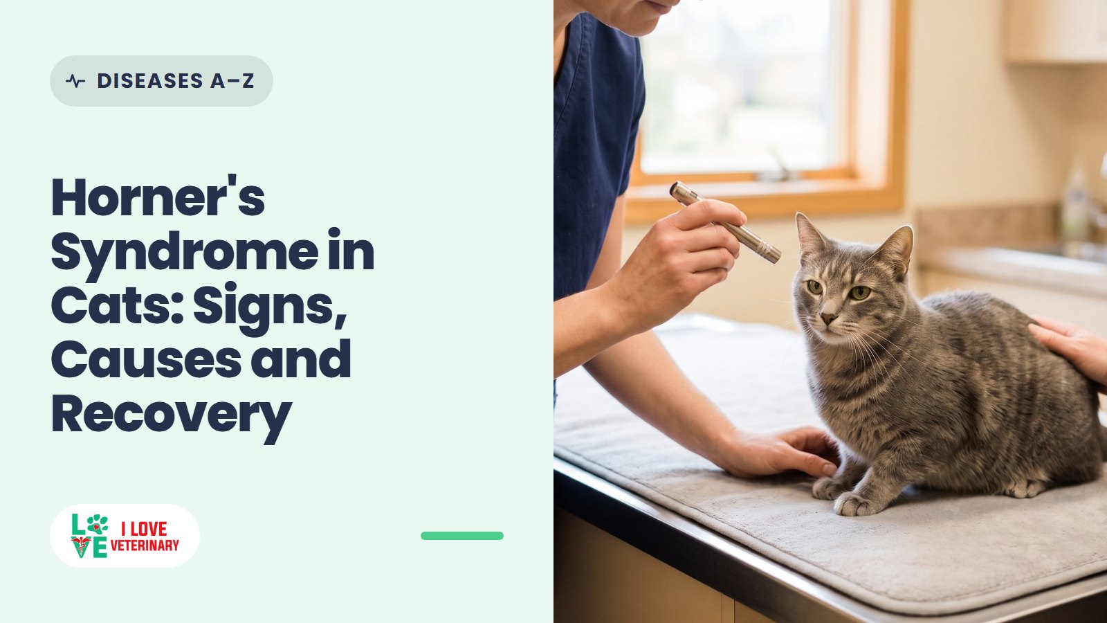
Horner's syndrome is a set of four eye signs on one side of a cat's face: a small pupil, a drooping upper eyelid, an eye that looks sunken, and a third eyelid that stays partly across the eye. It happens when the sympathetic nerve supply to that eye is interrupted somewhere along its route from the brain, down into the chest and back up past the ear.
The eye itself is not painful and your cat can still see. The signs are a clue that something has irritated or damaged that nerve. In cats, middle ear disease and injuries are common causes. Sometimes a full workup finds nothing at all. That is idiopathic Horner's syndrome, and many of those cases fade on their own over weeks to months.
If you notice these signs, book a visit with your veterinarian. Call now if your cat was recently hurt (a fall, a car, a fight), drags or can't use a front leg, is wobbly or tilting its head, or the eye is red, cloudy or painful.
The Four Signs of Horner's Syndrome in Cats
Horner's syndrome is not a disease on its own. It is a group of signs telling your vet that one nerve pathway is not working.
- Small pupil (miosis). The most common sign. It is easiest to see in a dim room, when the healthy pupil opens wide and the affected one can't. In bright light the two can look almost the same.
- Drooping upper eyelid (ptosis). The eye opening looks narrower, as if the cat is half squinting.
- Sunken eye (enophthalmos). The eyeball sits a little further back in the socket.
- Raised third eyelid. The pale third eyelid (nictitating membrane) slides partly across the inner corner of the eye. Unlike dogs, cats have a small muscle in the third eyelid that this same nerve supply controls.
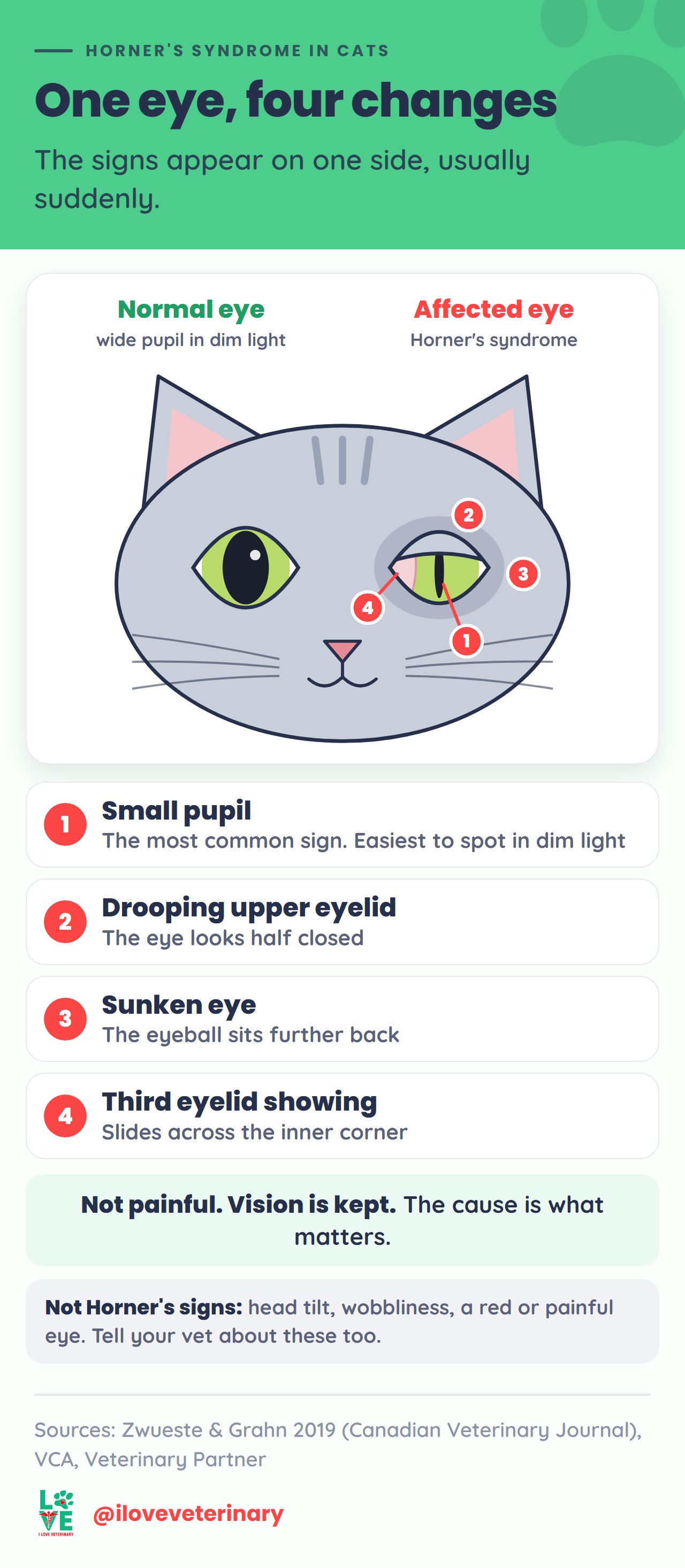
The signs usually appear suddenly and on one side. Now and then the ear on that side feels warmer or the nose looks pinker, because the same nerves also control blood vessels in the face.
What Is Not a Horner's Sign
These often turn up alongside Horner's syndrome but are not part of it. Tell your vet about them, because they point to the cause:
- Head tilt, circling, wobbliness or flicking eyes usually mean the inner ear, the balance centers of the brain or the spinal cord are involved too.
- A droopy lip or ear, or an eye that doesn't blink fully, suggests the facial nerve is affected as well, which often happens with middle ear disease.
- A red, cloudy, watery or painful eye points away from Horner's syndrome and toward inflammation inside the eye (uveitis) or a scratch or ulcer on its surface.
Does It Hurt? Can My Cat Still See?
Horner's syndrome itself is not painful, it does not take away vision, and the pupil still reacts to light. A third eyelid that sits far across the eye can block part of the view, and your vet can help with that.
The cause is another matter. A middle ear infection, an injury or a growth can be painful or serious, so the signs deserve a vet visit even when your cat seems fine.
Why It Happens: A Long Nerve Loop
The sympathetic nerves are the "fight or flight" system. For the eye, they widen the pupil, lift the eyelids and pull the third eyelid out of the way. When they stop working, the "rest and digest" side acts alone, and the four signs appear.
Their route to the eye is surprisingly long, in three segments:
- First segment (first order): from the brain down the spinal cord through the neck to the top of the chest.
- Second segment (second order): from the spinal cord at the top of the chest, through the chest, and back up the neck to a nerve junction just beside the middle ear.
- Third segment (third order): from beside the middle ear, through or next to the middle ear, to the eye.
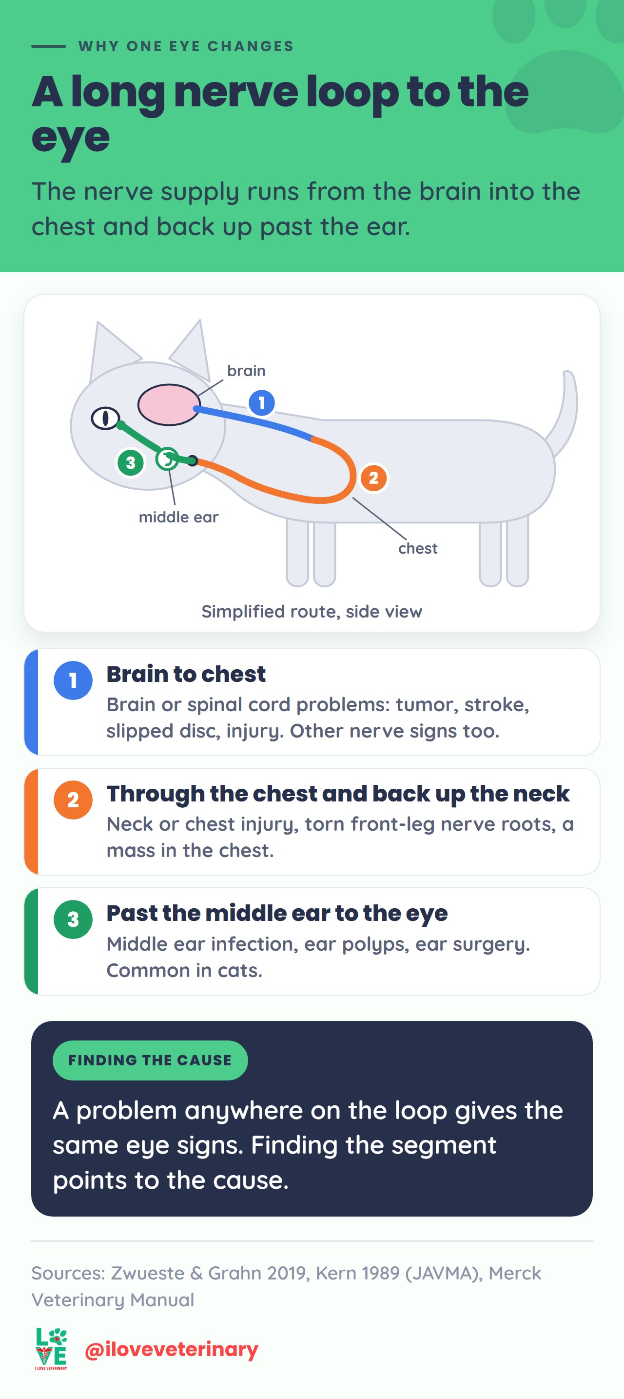
A problem anywhere on that loop causes the same four signs. Where it sits points to the likely cause, so finding the segment is the first job of the workup.
Common Causes in Cats
In a Cornell series of 100 dogs and cats with Horner's syndrome (26 of them cats), the main causes were injuries such as being hit by a car, torn nerve roots in a front leg, tumors in the brain or chest, and middle and inner ear infection. In the cats, ear infection stood out as a risk.
Third Segment: Middle Ear Problems
The nerve runs through or right next to the middle ear, so ear disease is a classic cause.
- Middle ear infection (otitis media). Horner's syndrome is one of the signs that tells a vet an ear infection has reached the middle ear. In a study of 196 cats with inner ear balance problems, it went with middle and inner ear infection.
- Inflammatory polyps. These benign growths are most common in young cats (about 3 months to 5 years old) and can grow into the middle ear. In one study, cats with polyps were much more likely to show Horner's syndrome.
- Ear canal tumors in middle-aged and older cats, once they reach the middle or inner ear.
- Ear procedures. Horner's syndrome is a common side effect of middle ear surgery in cats, and injury from ear canal cleaning has also been reported. Tell your vet about any eye change after an ear procedure.
Second Segment: Chest, Neck and Front Leg
- Injuries to the neck or chest, including bite wounds and road accidents.
- Torn nerve roots in a front leg (brachial plexus avulsion), usually after an accident. Horner's signs plus a limp front leg on the same side need a vet quickly.
- Masses in the chest, such as tumors pressing on the nerve as it passes through.
First Segment: Brain and Spinal Cord
Tumors, strokes, a slipped disc or a spinal cord injury in the neck can damage the first segment. These cats almost always have other nervous system signs too, such as weakness or wobbliness.
Idiopathic Horner's Syndrome in Cats
Idiopathic means no cause was found. Your vet reaches that diagnosis after the exam, ear check and imaging come back normal, though it may be suspected early when the signs came on suddenly and the rest of the exam is normal.
How often that happens in cats is not settled. An older Cornell study of 26 cats found no cause in about 4 in 10, while VCA's owner guide says a cause is almost always found in cats and truly idiopathic cases are very rare. Either way the workup is worth doing, because there is often an ear problem or an injury to treat. If nothing turns up, that is good news: idiopathic Horner's needs no specific treatment, and many cases improve on their own.
How Your Vet Finds the Cause
After an injury, your vet will stabilize your cat first. Then the usual steps are:
- History. Any fall, fight or accident, ear scratching or head shaking, recent ear cleaning or surgery, and when you first noticed the eye.
- Eye exam. To confirm the small pupil is the abnormal one and to rule out inflammation inside the eye or a corneal ulcer, which can look similar.
- Neurologic exam. Gait, reflexes and the facial nerves show whether the problem is likely in the brain, spinal cord, chest or ear.
- Ear exam. A look down the ear canal with an otoscope, sometimes under sedation, for infection, a bulging eardrum or a polyp. See what an otoscope can and can't show.
- Eye-drop test (in the clinic only). Your vet puts a diluted drop in both eyes and compares how quickly each pupil and eyelid responds. A fast response on the affected side points to the last segment, near the ear or behind the eye. It is more reliable once the signs are a couple of weeks old.
- Bloodwork and X-rays of the chest and neck, to check general health and look for a mass or injury.
- CT or MRI when the earlier steps don't find the cause, to see the middle ear and the whole nerve route.
Conditions That Look Like Horner's Syndrome
- Third eyelids showing in both eyes, without a small pupil. This often goes with general illness or dehydration. In Haw's syndrome both third eyelids come up, often with a stomach or gut upset, and settle within weeks.
- Facial nerve paralysis. A droopy face and an eye that can't blink, often with bad ear infections.
- Feline dysautonomia (Key-Gaskell syndrome). A rare, serious disease of the automatic nervous system. The pupils are wide and don't react, the eyelids droop or the third eyelids show, tears dry up, and cats often have constipation, vomiting or trouble swallowing. Here the pupils are large, not small.
- Eye inflammation or a corneal ulcer. Can cause a small pupil, but the eye is usually red, watery or held shut. See our guide to why cats get watery eyes.
Treatment
Treatment is aimed at the cause, not the eye. Your vet treats a middle ear infection (some cats need ear surgery), removes polyps, and plans care for an injury or tumor based on what they find. Idiopathic Horner's needs no specific treatment, just rechecks.
The eye signs themselves often need nothing. If the third eyelid gets in the way, your vet may prescribe drops that pull it back for a while (they can irritate the eye). If your cat can't blink properly or the eye is dry, your vet may add a lubricating eye product to protect the cornea.
Don't put any drops or ointment in your cat's eye, or clean deep into the ear, unless your vet has told you to.
Recovery and Outlook
The outlook depends on the cause.
- When no cause is found, or the injury was minor, many cases improve on their own. Most recovery figures come from dogs or mixed groups of dogs and cats. In one study of both, full recovery took about 8 weeks on average (7.7 weeks, all causes together), and idiopathic cases in dogs improve over 4 to 15 weeks. For a cat, expect weeks to a few months and keep your rechecks.
- Some animals keep a slightly smaller pupil after the other signs fade. That is cosmetic.
- With a middle ear infection, the eye signs can resolve once the infection is treated.
- After middle ear surgery, Horner's syndrome followed about two thirds of the operations in a study of 282 cats. Some cats kept it permanently, more often those that had it before surgery.
- When the front leg on the same side is completely paralyzed, the nerve roots are usually torn and the leg is unlikely to recover.
- With tumors, the signs are more likely to be permanent.
When to Call Your Vet
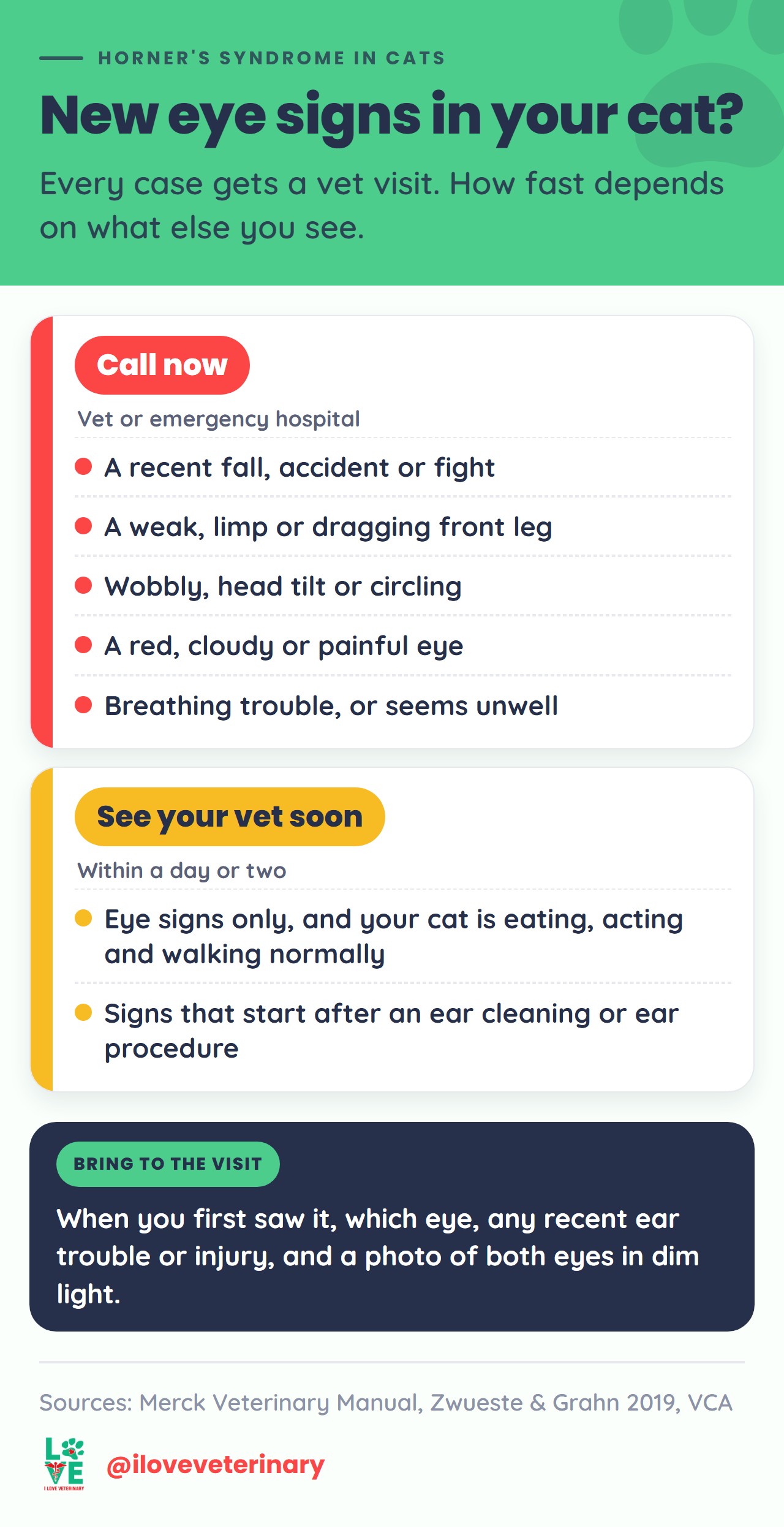
Call now if Horner's signs come with any of these:
- A recent fall, road accident or fight
- A front leg that is weak, limp or dragging
- Wobbliness, a head tilt, circling or flicking eyes
- A red, cloudy or painful eye, or your cat keeps the eye shut
- Trouble breathing, or your cat seems unwell
See your vet soon (within a day or two) if your cat has new Horner's signs but is otherwise eating, acting and walking normally, or if the signs appear after an ear cleaning or ear procedure.
Before the visit, note when you first saw the change, which eye it is, and any recent ear trouble, ear procedure or possible injury. A photo of both eyes in room light and one in dimmer light helps your vet compare the pupils.
Frequently Asked Questions
Can Horner's syndrome affect both eyes?
It usually affects one side. Both sides are possible but uncommon; raised third eyelids in both eyes more often mean general illness or dysautonomia.
Will it come back?
Repeat episodes are unusual in dogs with idiopathic Horner's. There is little data for cats, so mention any new eye change to your vet.
Originally written by Jaclyn. Updated by the I Love Veterinary editorial team.
Sources
- Zwueste DM, Grahn BH: A review of Horner's syndrome in small animals (Canadian Veterinary Journal, 2019)
- Kern TJ and colleagues: Horner's syndrome in dogs and cats, 100 cases (JAVMA, 1989)
- Morgan RV, Zanotti SW: Horner's syndrome in dogs and cats, 49 cases (JAVMA, 1989)
- Wainberg SH and colleagues: Complications and outcome after ventral bulla osteotomy in 282 cats (JAVMA, 2019)
- Spivack RE and colleagues: Complications after total ear canal ablation in dogs and cats (JAAHA, 2013)
- Grapes NJ and colleagues: Clinical reasoning in feline vestibular syndrome (Journal of Feline Medicine and Surgery, 2021)
- Caldero Carrete J and colleagues: Peripheral vestibular syndrome in cats, 196 cases (Veterinary Record, 2025)
- Merck Veterinary Manual: Otitis Media and Interna in Animals
- Merck Veterinary Manual: Overview of Tumors of the Ear in Small Animals
- Merck Veterinary Manual: Inflammatory Polyps in Cats
- Merck Veterinary Manual: Leg Paralysis in Cats
- Merck Veterinary Manual: Dysautonomia in Cats
- Veterinary Partner (VIN): Horner's Syndrome in Cats and Dogs
- VCA Animal Hospitals: Horner's Syndrome in Cats
- Mar Vista Animal Medical Center: Horner's Syndrome
This article is for general information and is not a substitute for advice from your veterinarian.