
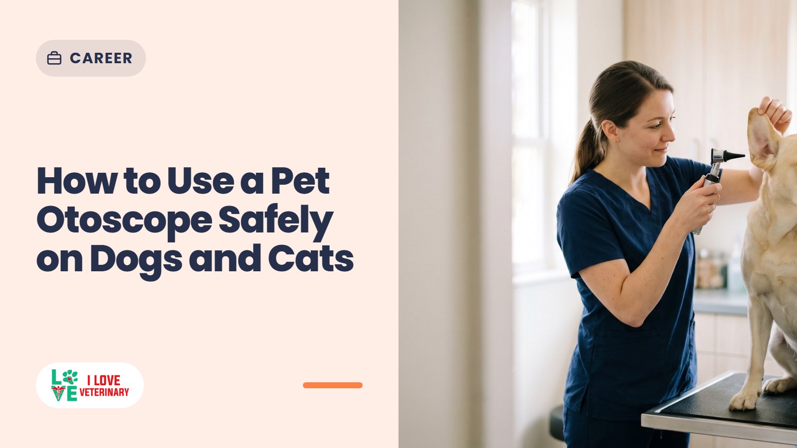
A pet otoscope shines a bright, magnified light into your dog's or cat's ear, so you can see the ear flap, the canal opening and the first part of the canal. Past that point a home check stops being safe. A dog's ear canal bends partway down, the eardrum sits around that corner, and reaching it without hurting the animal takes training, good restraint and sometimes sedation.
So the rule for owners is simple: look, don't probe. Use it to spot redness, discharge or something stuck near the opening, then let your veterinarian go deeper. Vet students and new techs: the full technique is in the last section.

Ear problems are common in dogs and cats. Head shaking, scratching, a red or smelly ear and a dark or yellowish discharge are the classic signs. Floppy, hairy or narrow ears, allergies, parasites such as ear mites, and grass seeds can all set things off.

What an Otoscope Is
An otoscope (also called an auriscope) is a handheld light and magnifier for looking into the ear canal. A standard one has:
- A handle that holds the battery.
- A head with a light and a low-power magnifying lens.
- A speculum, the cone-shaped tip that sits in the ear. Cones are usually disposable or washable and come in different widths.
On some models the lens comes off so the vet can pass small instruments through the cone. A video otoscope puts a camera at the tip and shows the canal on a screen, with more magnification, better light and recording. Some consumer otoscopes connect to a phone and take photos. Those are handy to show your vet, but they don't replace the exam.

Why a Dog's Ear Canal Bends (and Why You Lift the Ear Flap)
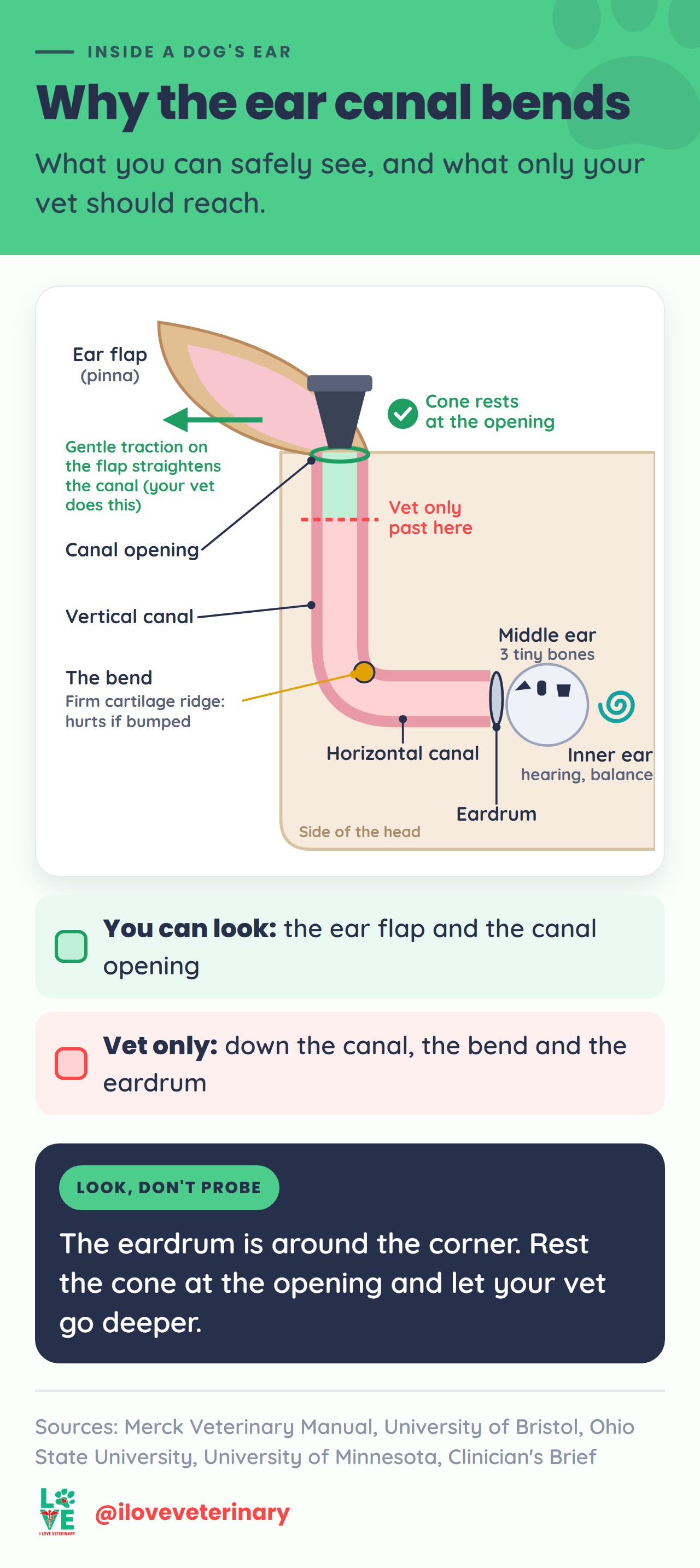
A dog's ear canal is much longer than ours, and it isn't straight. It runs down the side of the head as the vertical canal, then turns sharply inward as the horizontal canal. The eardrum (tympanic membrane) sits at the far end of the horizontal part. Behind it are the middle ear, with its three tiny hearing bones, and the inner ear, which handles hearing and balance.
That bend is why examiners hold the ear flap (pinna) and apply gentle traction to it. Traction straightens the canal so the cone can follow it instead of pressing into the wall. The bend also hides a firm ridge of cartilage that hurts if the cone bumps it. Cats are examined with the same technique.
For owners, that settles it. Everything you can safely see sits near the opening of the vertical canal, and the eardrum is around the corner, out of reach for a home check.
Choosing the Right Speculum Size
- Match the cone to the ear. A diameter that suits the canal opening lets you keep the canal centered instead of scraping its sensitive wall.
- Clean and dry before every use.
- One cone per ear. Use a fresh or disinfected cone for the second ear so you don't carry debris or infection across.
If no cone in the set sits comfortably at the opening, don't force one.
How to Look in Your Pet's Ears Safely
This is our suggested home routine. It deliberately stops at the canal opening.
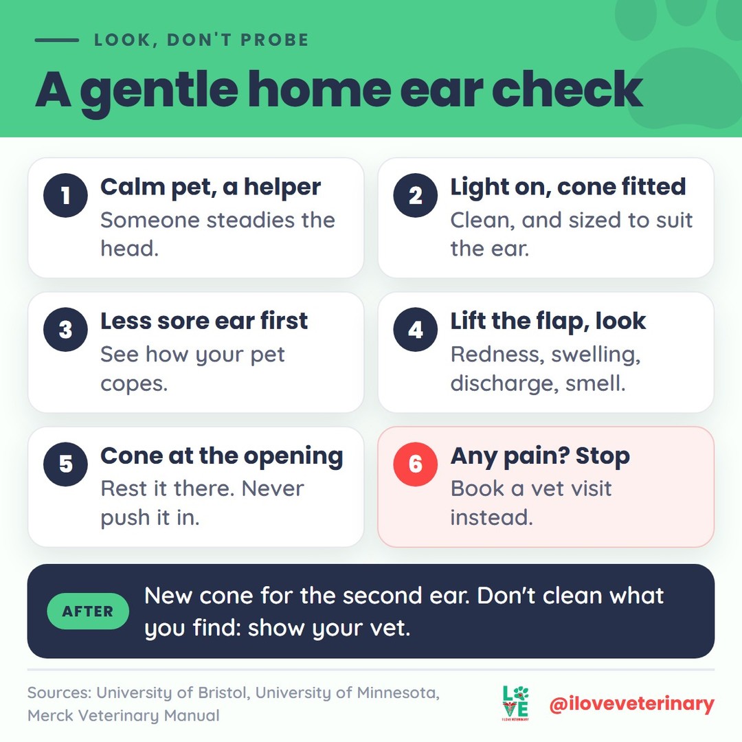
- Pick a calm moment. Have a second person gently steady your pet's head, and use treats to keep things relaxed.
- Turn the light on and check the cone is fitted firmly.
- Start with the ear that seems less sore. You'll see how your pet tolerates handling before you touch the worse ear.
- Lift the ear flap gently and look at its inner surface and the canal opening for redness, swelling, crusts, scratches or discharge. Sniff too. An infected ear often smells.
- Rest the cone at the opening, without pushing it in, and look into the first part of the canal. Keep looking through the lens the whole time so you know where the tip is.
- Stop at the first sign of pain. Pulling away, yelping, growling or head shaking means stop. Ear pain can make even the friendliest dog snap, so book a vet visit instead of trying again.
- Switch to a clean cone for the other ear, then clean or throw away the cones and turn the otoscope off.

What Normal and Problem Ears Look Like
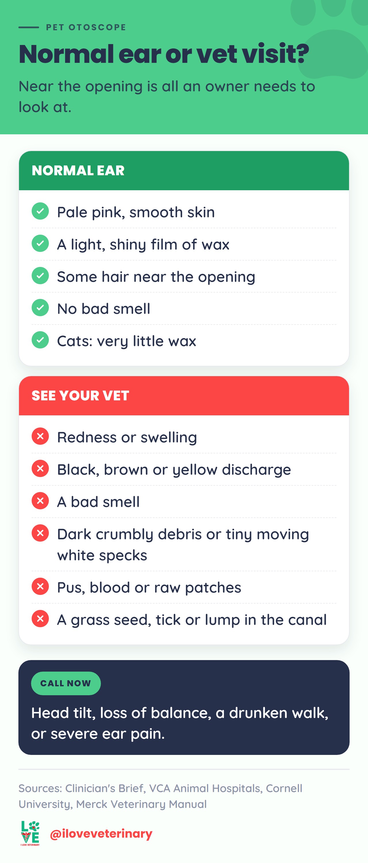
A healthy canal is smooth, pale pink and slightly glistening, with a thin coat of wax and some hair near the opening. Cats normally make very little wax.
What you see | What it can mean | What to do |
|---|---|---|
Pale pink, smooth skin, a light shiny film of wax, some hairs | A normal ear | Leave it alone. Healthy ears clean themselves |
Redness, swelling, or a canal that looks narrower than usual | Inflammation of the ear canal (otitis externa), often linked to allergies in dogs | See your vet soon |
Black, brown or yellow discharge, or a bad smell | Bacterial or yeast infection, or ear mites. They look alike | See your vet soon. A microscope check of the debris tells them apart |
Dark, crumbly debris, sometimes with tiny white moving specks | Possible ear mites, especially in kittens and puppies | See your vet soon |
Pus, blood, or raw, ulcerated patches | Severe infection, or a growth that has ulcerated | See your vet soon |
A grass seed, foxtail, tick or other object in the canal | A foreign body | See your vet as soon as possible. Don't pull it out: vets flush it out or remove it with forceps, often under sedation |
A lump or growth in the canal | Polyp, cyst or tumor (only a biopsy tells which) | See your vet soon |
Head tilt, loss of balance, a drunken walk, or scratching hard enough to wound the face | The problem may have reached the middle or inner ear | Call now |
Discharge color is only a first hint. Vets confirm the cause by checking a sample under the microscope.
Why You Shouldn't Push Deep or Clean What You See
It's tempting to chase the gunk you spot. Please don't. Here's why:
- The eardrum has to be checked before anything goes into the ear. If it is torn, some ear medications can cause hearing loss. Only an exam that reaches the eardrum confirms it is intact, and in a sore, swollen or dirty canal even vets often can't see it until the ear settles.
- Aggressive cleaning can rupture the eardrum. The Merck Veterinary Manual lists eardrum rupture from aggressive cleaning among the causes vets consider when a dog suddenly develops middle ear disease.
- Normal wax is not a problem to fix. Skin in the canal slowly moves outward and carries wax with it. A peer-reviewed Clinician's Brief guide advises against ear cleaners for small amounts of wax in normal ears, and Merck lists overcleaning as a cause of ear inflammation.
- Your vet needs the evidence. Vets take a swab of the debris for the microscope before any cleaning. Clean first and the clue is gone.
The same goes for anything you find. Grass awns such as foxtails can work deep enough to cause middle ear disease, and a sore ear canal is no place for tweezers.

When to Call Your Vet
Call now if your pet:
- Tilts its head, loses its balance or walks as if drunk.
- Is in obvious, severe ear pain or scratches so hard it wounds its face.
- Suddenly starts shaking its head or pawing at one ear after being in dry grass. A foxtail in the ear needs a vet as soon as possible.
See your vet soon for:
- Head shaking, scratching or rubbing an ear.
- Redness, swelling, odor or any discharge.
- Anything stuck in the canal, or a lump.
- A soft, puffy swelling of the ear flap. It can be an aural hematoma, which needs a vet.
If the ears look fine and your pet is comfortable, leave them be and mention anything you noticed at the next checkup.
For Vet Students and New Techs: The Full Otoscopic Exam
The complete exam goes all the way to the eardrum, and good technique is what keeps it from hurting.

- Prepare. Fit the head to the handle, check the light, and choose a clean, dry cone of the right diameter. Consider a muzzle, because even friendly dogs may bite when an ear hurts. Warn the owner that the exam can be uncomfortable.
- Start with the less affected ear. Lift the pinna, examine its concave surface and the canal opening, and smell the ear.
- Enter the vertical canal. Introduce the tip at the opening and advance slowly while looking through the scope. Keeping the lumen centered stops the cone scraping the canal wall.
- Turn the corner. As the canal narrows and bends, apply gentle traction to the pinna to straighten it. Sources describe the direction differently (up and toward you in one vet-school handbook, outward and downward in a clinical guide). Either way, the aim is to line the canal up so the cone passes under the cartilage ridge (Noxon's ridge) instead of hitting it. The speculum ends up roughly horizontal, at about 90 degrees to the skull.
- Assess the canal. Note wax and exudate, foreign bodies such as grass awns and ticks, live ear mites (white specks moving on dark debris), redness, ulceration, masses, and how narrowed the canal is. The horizontal canal normally has far less hair than the vertical canal.
- Assess the eardrum. A normal pars tensa is thin, pale gray and translucent. Above it, the pars flaccida is the fleshier, vascular part and can bulge in some dogs. The handle of the malleus shows through the pars tensa: a gentle forward-pointing hook in dogs, thinner and straight in cats. Mucus in the external canal suggests a hole in the eardrum, because the canal itself makes none.
- Finish. Withdraw slowly, wipe the cone, and use a clean cone for the second ear. Turn the otoscope off and clean or disinfect the specula.
An ear that is too painful, swollen or full of exudate to examine gets either sedation or anesthesia the same day, or treatment for the swelling and pain first and a recheck once the canal opens. Sample exudate for cytology before any cleaning or flushing. A video otoscope makes the eardrum easier to assess and records findings for the chart and the client.
Sources
- Merck Veterinary Manual: Otitis Externa in Animals
- Merck Veterinary Manual: Otitis Media and Interna in Animals
- Merck Veterinary Manual (Dog Owners): Ear Structure and Function in Dogs
- Merck Veterinary Manual (Cat Owners): Ear Structure and Function in Cats
- VCA Animal Hospitals: Ear Infections in Dogs
- VCA Animal Hospitals: Ear Infections in Cats
- Cornell University College of Veterinary Medicine: Itchy Ear Problems
- UC Davis School of Veterinary Medicine: Foxtails and Dogs
- Clinician's Brief: Examination of the Ear
- Clinician's Brief: Examining the Ear: Otoscopy in Dogs and Cats
- University of Bristol Veterinary School: Otoscopic Examination (clinical skills booklet)
- The Ohio State University: Veterinary Clinical Skills Handbook, Otoscopic Examination
- University of Minnesota: Otoscopic Exam in Dogs and Cats (Veterinary Clinical Skills Compendium)
This article is for general information and is not a substitute for advice from your veterinarian.