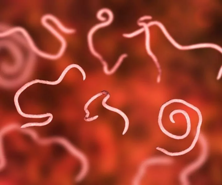
Entomopathogenic Nematodes are considered to be the most numerous multi-cell organisms on the planet. The phylum Nemata contains over 20.000 classified species. Structurally they are simple organisms.

Adult forms of the organism are comprised of around 1.000 cells, most of them as a part of the digestive system. Nematodes possess digestive, excretory, reproductive and nervous systems and lack circulatory and respiratory systems.
The ‘tube within a tube’ phenomena refer to the long alimentary canal extending from the mouth of the parasite to the anus (anterior end). They can range from 0.3 mm to over eight meters in size.
We will take you back to those parasitology classes by describing the most relevant Nematode species in the animal kingdom. For the purpose of simplifying the article and making it easy to use we divided them into two categories, intestinal and non-intestinal, and subsequently in genuses.
Intestinal Nematodes
Ascarids (Roundworms)
Toxascaris leonina is a parasite whose infestations usually result in asymptomatic illness. They can be found everywhere in Europe and the final hosts are dogs, cats, and foxes.
The infestation occurs by ingestion of embryonated eggs present in the soil or paratenic hosts larvae. For cats, there is a pre-patent period of 13 weeks, eight weeks in dogs, and a patent period of four to six months both for dogs and cats.
Toxascaris leonina eggs can be detected using the flotation method using between three to five grams of feces. There is a risk of zoonosis, especially in children.
Toxocara cati can be found in cats as final hosts and the infestation is due to ingestion of embryonated eggs present in soil and larvae from milk and paratenic hosts.
The infestations can result in asymptomatic cases or can cause intestinal symptoms due to intestinal blockage and intussusceptions. Young kittens suffer from cachexia, occasional pneumonia and are presented with pot-like bellies.
The Pre-patent period is six weeks and the patent period is between four to six months. Even though they can be transmitted to humans, the risk is lower than Toxocara canis. An identical flotation method is used for the detection of eggs.
Hookworms
Ancylostoma caninum, also known as dog hookworm can cause acute or chronic illness with diarrhea (sometimes bloody), anemia, and weight loss in dogs and foxes.
The ingestion of third-stage larvae (L3) from the environment or milk/paratenic hosts larvae is one way of transmission, but percutaneous infestation has also been reported. Detection of eggs is performed by using the standard flotation method.
Ancylostoma tubaeforme infects cats resulting in chronic or acute signs of diarrhea, bloody diarrhea, anemia, and weight loss. The parasite can be predominantly found in Europe and isn’t a threat to human health.
Uncinaria stenocephala has cats, dogs, and foxes as final hosts. Ingestion of embryonated eggs from soil and larvae from paratenic hosts results in a disease with acute or chronic gastrointestinal clinical signs (diarrhea, weight loss).
The pre-patent period is three to four weeks and the patent period varies depending on the immunological status of the final host.
Whipworms
The last representative of intestinal nematodes is Trichuris vulpis. Mild infestations are usually asymptomatic, but heavy cases can cause illness manifested with diarrhea, weight loss, and anemia.
Dogs that ingest embryonated eggs present in the environment are the final hosts. The pre-patent period lasts for about eight weeks followed by long a 18-month patent period.
Non-intestinal Nematodes
Heartworms
Dogs and foxes as final hosts for Angiostrongylus vasorum get infected by ingesting infective larvae present within mollusks or paratenic hosts. The patent period of up to five years follows a pre-patent period of 40-49 days.
Initially, the disease is asymptomatic after which generalized respiratory signs are apparent with cough, tachypnoea, and dyspnoea in most cases. Other pathologies include coagulopathy and neurological signs. Sometimes sudden death occurs without initial respiratory signs.
A serological test for detecting Angiostrongylus vasorum is commercially available. Other diagnostic methods used are the Baermann method for detection of live larvae using at least four grams of feces and microscopic larvae detection from bronchial lavage.
Dirofilaria immitis occurs rarely in cats and the transmission of the infective 3rd stage larvae is via an intermediate host (mosquito). The pre-patent period lasts approximately eight months and most of the time the infections are asymptomatic.
Once the worms reach the heart, acute respiratory and circulatory symptoms appear with coughing, tachycardia, and tachypnoea. The definitive diagnosis of heartworm can only be done by a combination of serological tests, hematological tests with echocardiography, and thoracic radiography.
Even though reported, human infections rarely occur.
In dogs, Dirofilaria immitis has a pre-patent period of 120-180 days and a patent period of several years. Mild infections result in asymptomatic disease, but severe infections may pose a serious threat to the dog’s life and can prove to be fatal.
Between five to seven months after infection, the initial clinical signs appear coughing, dyspnoea, and lack of stamina. The disease can develop into a chronic form manifested through ‘Caval syndrome’, tachycardia, tachypnoea, and frequent coughing.
Detection after concentration of microfilaria using Difil or Knott’s Test 180 days after infections shows great diagnostic results. Serological antigen detection 5 months after infection can potentially have 100% sensitivity if at least 1 female worm is present.
Read more about Heartworm resistance here.
Lungworms
Aelurostrongylus abstrasus is generally prevalent in stray cats (final hosts) that get infected by ingesting an intermediate host. Most of the cats show no signs of illness, and the clinically apparent cases experience exercise intolerance and respiratory symptoms.
The patent period is several years and the pre-patent period lasts between seven to nine weeks. Bronchial lavage larvae microscopic detection and the Baermann method are used as diagnostic tools.
Capilaria spp. can cause asymptomatic to fatal diseases in dogs, cats, and foxes depending on the specific parasite species and quantity in the organism. The infection occurs by ingestion of infective larvae from the environment followed by four weeks of pre-patent and 10-11 months of patent period.
Capilaria hepatica causes hepatic lesions and usually has fatal outcomes and Capilaria philippinensis damages the small intestine resulting in fatal enteropathy. The infections are generally discovered during routine autopsies. C. hepatica, C. philippinensis, and C. aerophila can infect people.
Final hosts for Crenosoma vulpis (fox lungworm) are dogs and foxes that get infected by ingesting larvae present in mollusks and paratenic hosts. The parasite generally affects the respiratory system with corresponsive clinical signs. Microscopic evaluation of bronchial lavage and Baermann methods are used for detecting larvae.
Filaroides hirthi infects dogs by an unknown route of transmission and potentially inflicts respiratory symptoms (coughing, exercise intolerance). The pre-patent period lasts 10-18 weeks and there is a lack of information regarding the patent period.
Diagnostic methods for the detection of larvae are the Baermann method and bronchial lavage microscopic evaluation.
Oslerus osleri is directly transmitted orally from bitches to pups in dogs and foxes. The usual respiratory signs are apparent and besides bronchial lavage and feces evaluation, endoscopy and radiography are useful for diagnosing.
Subcutaneous worms, oesophageal worms and threadworms
Dirofilaria repens infects cats, dogs, and other carnivores and is transmitted via mosquitoes (cutaneous infection). The pre-patent period is up to 34 weeks followed by several years of patent period.
Even though mostly asymptomatic, cutaneous lesions sometimes occur. The diagnostic methods are the same as for the previous Dirofilaria species using a two to four ml EDTA blood sample. There is a zoonotic risk and infections result in subcutaneous nodules in the conjunctiva.
Spirocera lupi (oesophageal worm) infections can cause alimentary disruptions in dogs, cats, foxes, wild dogs, and wild cats. Many cases are without clinical signs and the ones clinically apparent exhibit vomiting (with worms in the content) and difficulty swallowing.
Endoscopic and radiographic images show granulomatous lesions inside the esophagus.
Infections with Strongyloides stercoralis in dogs, cats, and humans as final hosts, resulting in bloody diarrhea, severe dehydration, and sometimes death. The route of transmission is by ingestion of embryonated eggs from fur and soil, ingestion of larvae present in milk and paratenic hosts, and also vertically.
In people, the parasite causes several forms of the disease: chronic intestinal syndrome, subcutaneous lesions, mild transient form, and neurological illness. The pre-patent period is short (nine days) and the patent period ranges between three to 15 months.
Eyeworms
Thelazia callipaeda is transmitted onto dogs and cats via arthropod vector. The patent period lasting few months to a couple of years follows a pre-patent period of about three weeks.
The infections result in epiphora and blepharospasms. The material used for the detection of adult forms of the parasite and larval stages is tear film taken from the surface of the conjunctiva.
