
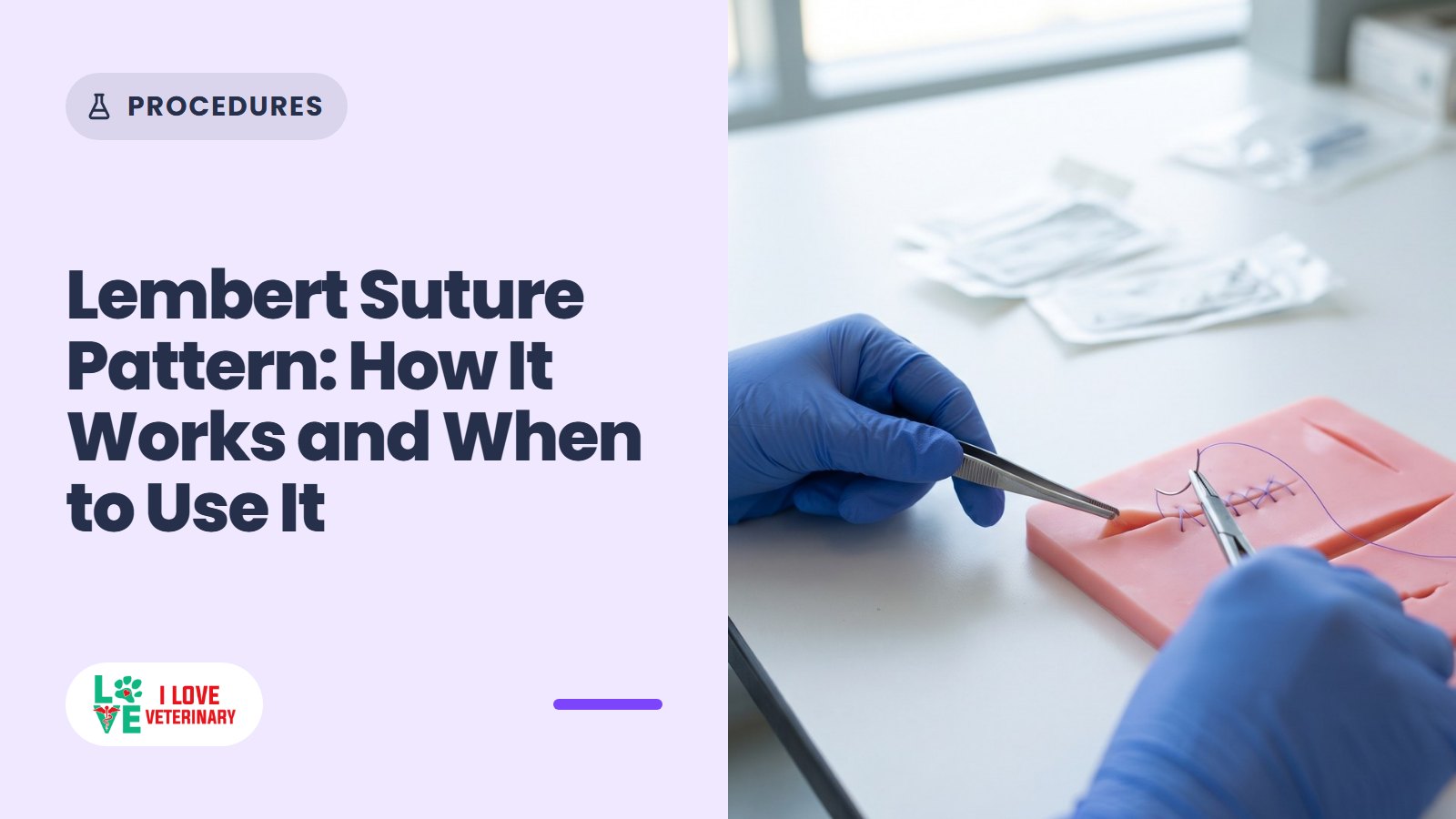
The Lembert suture pattern is an inverting stitch for hollow organs. In veterinary surgery it is often placed on the stomach, urinary bladder and uterus, where it folds the cut edges inward so the outer surfaces (serosa) meet and the suture line is buried. That helps keep the organ's contents in and leaves little suture exposed on the outside.
The needle never passes all the way through the wall, so the suture stays out of the lumen. In dogs and cats the Lembert is usually the second, "oversew" layer laid over a first closure. You can watch the full pattern in this short Lembert suture pattern video from LMU's College of Veterinary Medicine.
How the Lembert pattern works
A hollow organ wall has four layers: serosa on the outside, then muscularis, submucosa, and the mucosa lining the lumen. The submucosa is the strength layer, and Guelph's surgery text says it must be included in the bites of an intestinal closure. WCVM's surgery lab describes the Lembert bite the same way: through the serosa and muscularis and into the submucosa, but not through the mucosa, so the needle does not enter the lumen (WCVM).
The bites run perpendicular to the incision. As each one tightens, the edges roll inward and the serosa on one side presses against the serosa on the other. Done well over a first layer, the inverted tissue covers that first line so completely that only the knots of the Lembert remain visible (Guelph Veterinary Surgery Online).
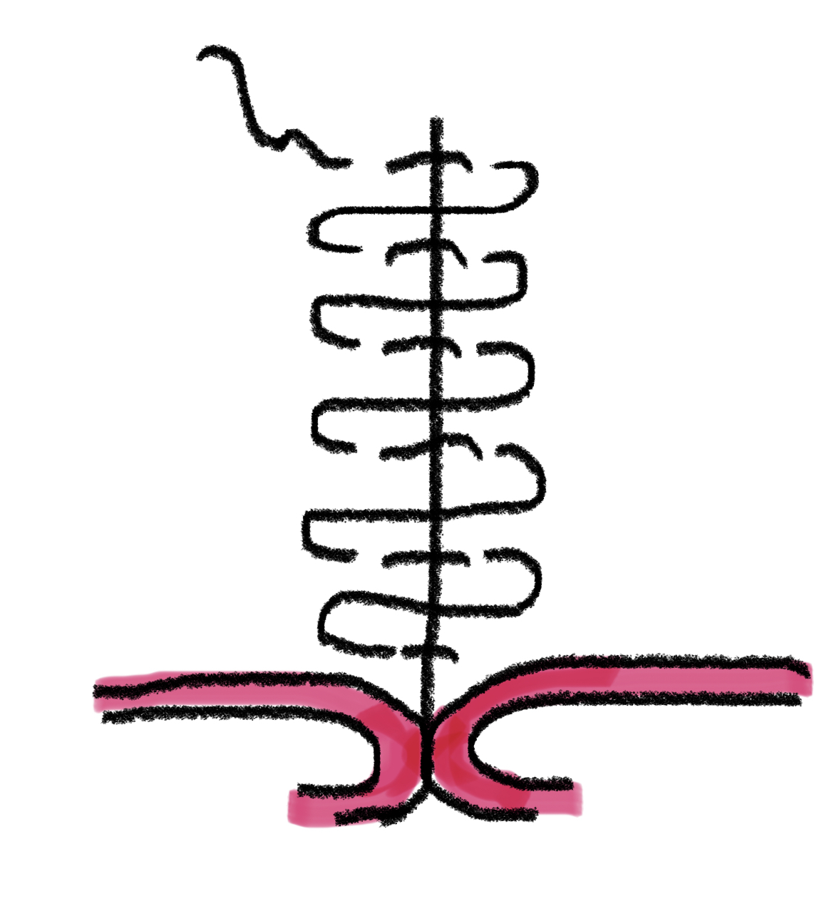
That inversion is the point of the pattern and also its cost. Every fold of tissue that turns in takes up room inside the organ, and the pattern affects the local blood supply more than a simple continuous line does. Both trade-offs shape where surgeons use it.
How a continuous Lembert is placed, step by step
The University of Guelph's surgery teaching text describes the continuous Lembert in this sequence. Its shorthand for each bite is "through the serosa and muscularis"; in WCVM's terms that bite reaches into the submucosa without entering the mucosa.
- The line starts at one end of the incision with a partial-thickness simple throw across it, which is tied.
- On one side, the needle goes in perpendicular to the incision about 8-10 mm from the edge and comes out on the same side about 4 mm from the edge.
- The suture crosses the incision. The needle enters the opposite side about 4 mm from the edge, again perpendicular, and comes out 8-10 mm from the edge on that same side.
- The line advances along the incision, as in a simple continuous pattern, and the sequence repeats: far to near on one side, near to far on the other.
- At the end of the incision the suture is knotted.
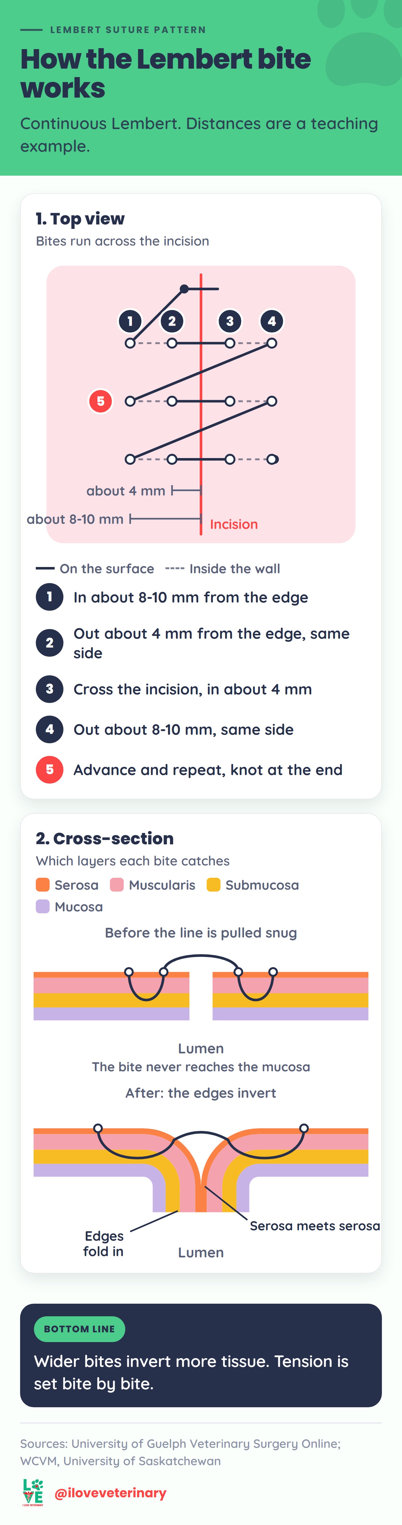
The 8-10 mm and 4 mm figures are a teaching example, not a fixed rule. WCVM adds the principle behind them: the farther from the edge the bites are placed, the more tissue gets inverted, so bite width controls how much wall folds in. Its other rule is about tension. It is set bite by bite as the line goes, because it is hard to readjust once the pattern is finished.
Where the Lembert pattern is used
Dogs and cats: usually the second layer
In small animal surgery the Lembert most often appears as the oversew layer in a two-layer closure. The first layer apposes the edges, usually with a simple continuous pattern, and the Lembert (or a Cushing) on top inverts and buries it. Guelph's surgery text uses it this way in three common procedures:
- Gastrotomy (an incision into the stomach, often to remove a foreign body): a full-thickness simple continuous layer, then a Lembert or Cushing oversew. Two inverting lines are another option.
- Cystotomy (an incision into the bladder, often to remove stones like those seen in cats with lower urinary tract disease): a simple continuous appositional layer, then a Lembert or Cushing over the seromuscular layer. A single continuous layer is an alternative and is preferred when the bladder wall is thick.
- Hysterotomy during a C-section: a simple continuous layer, sometimes followed by a Cushing or Lembert.
Large animals: intestine, bladder and uterus
Inverting patterns play a bigger role in horses and cattle. University of Minnesota large animal surgery notes list them for closing the intestine, bladder and uterus, because they prevent leakage and keep suture off the outer surface, which lowers the risk of adhesions. Small animals are less prone to adhesions, so other patterns may be used in them (UMN Large Animal Surgery).
The Lembert can also stand alone as a single layer. In a 2006 study from UC Davis, end-to-end anastomoses (rejoined cut ends) of jejunum from 12 horses were closed either with a single continuous Lembert or with a simple continuous layer oversewn with a Cushing. The one-layer Lembert was faster to place, left a larger opening, and burst at the same pressure. Both techniques narrowed the lumen compared with normal tissue. This was a laboratory study on equine intestine, so it supports the single Lembert as a reasonable option for equine jejunum, not as a better pattern everywhere.
Where the Lembert is usually not the first choice
In dogs and cats, the small intestine is normally closed with appositional sutures, not inverting ones. Guelph's text describes an appositional simple interrupted pattern as the typical enterotomy and anastomosis closure. A 2019 review explains why: approximating (appositional) patterns preserve luminal diameter, heal well, and match inverting patterns for bursting strength after the first 24 hours (Ellison and colleagues, Veterinary Surgery). An inverting pattern folds tissue straight into the lumen, so a pattern that works well on a stomach or a horse's bowel is not the default for a dog or cat enterotomy.
Lembert vs Cushing vs Connell
All three are continuous inverting patterns used on hollow organs. The differences come down to which way the bites run and whether the needle enters the lumen.
Pattern | Bite direction | Layers caught | Enters the lumen? | Memory hook |
|---|---|---|---|---|
Lembert | Perpendicular to the incision | Serosa, muscularis, submucosa | No | LemberT ends in T, for transverse |
Cushing | Parallel to the incision | Serosa, muscularis, submucosa | No | No "L" in Cushing: no lumen |
Connell | Parallel to the incision | Full thickness | Yes | ConnelL has the L: it goes into the lumen |
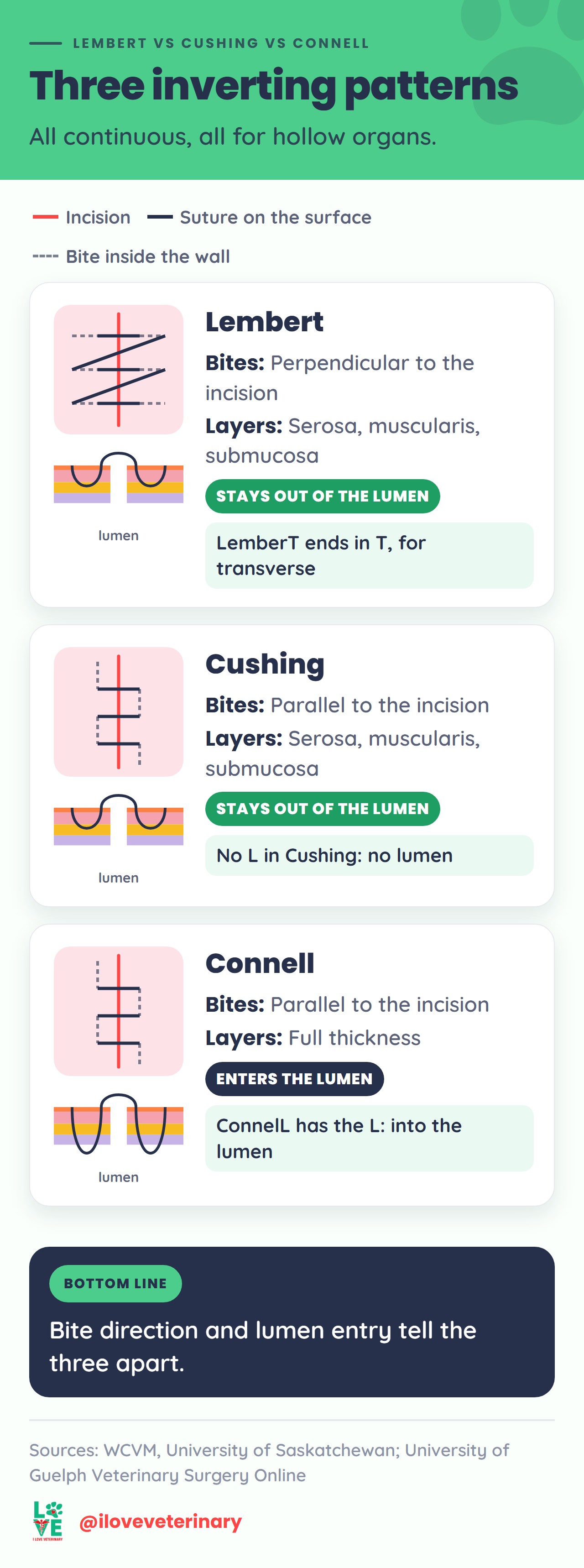
The memory hooks come from WCVM's surgery lab. Because the Connell passes into the lumen, it is not the pattern to pick when the goal is to keep suture out of the organ's contents. For more on the other two, read our guide to the Cushing and Connell suture patterns.
Who was Antoine Lembert?
Antoine Lembert (1802-1851) published Mémoire sur l'entéroraphie in 1826, describing a way to suture the intestine so that serous surface met serous surface (Garrison-Morton entry). His key step was leaving the mucosa out of the stitch and passing it through the serous and muscular coats only.

The submucosa came later. By 1895, an account of intestinal anastomosis in The Hospital credited Halsted with insisting that some submucosa be included in the bite, because sutures through the serosa and muscularis alone tend to cut out (Bidwell, via PubMed Central). The modern Lembert carries both ideas: serosa against serosa, and a bite deep enough to catch the submucosa.
Lembert's name also survives in the Czerny-Lembert closure, a two-layer hand-sewn intestinal anastomosis with a Lembert row on the outside. A 1992 study in pigs used it as the hand-sewn comparison against circular-stapled colorectal anastomoses and found the two healed in a similar way.
How the Lembert pattern is classified
Suture patterns are usually described by a few properties. For the Lembert:
- Continuous or interrupted: the teaching texts cited here describe the continuous form.
- Inverting, appositional or everting: always inverting.
- Depth: partial thickness. The bites stay out of the lumen.
- Bite direction: perpendicular to the incision.
- Layers: a single-layer closure on its own, or the second layer of a two-layer closure.
Suture material and needle
The surgeon chooses the material for each organ and patient. For the gastrointestinal tract, Guelph's text recommends a monofilament absorbable suture on a taper or taper-cut needle, advises against chromic gut, and notes that nonabsorbable monofilament is generally unnecessary. As typical examples it gives 3-0 (2-0 in very large dogs) for a gastrotomy and 3-0 or 2-0 for the uterus after a C-section. For the bladder it favors a rapidly absorbable monofilament and again rules out chromic gut, which loses strength very quickly in urine.
For the full textbook treatment of suture patterns and abdominal surgery, our list of the best veterinary books and study material is a good place to start.
Originally written by Helen Roberts. Updated by the I Love Veterinary editorial team.
Sources
- University of Guelph, Veterinary Surgery Online: Lembert Pattern
- University of Guelph, Veterinary Surgery Online: Cushing Pattern
- University of Guelph, Veterinary Surgery Online: Gastrotomy Closure
- University of Guelph, Veterinary Surgery Online: Cystotomy Closure
- University of Guelph, Veterinary Surgery Online: Close the Uterus in 2 Layers
- University of Guelph, Veterinary Surgery Online: Intestinal Apposition and Closure
- University of Minnesota, Large Animal Surgery: Inverting Suture Patterns
- Western College of Veterinary Medicine, University of Saskatchewan: Continuous Patterns
- Nieto JE et al.: One-layer (continuous Lembert) vs two-layer anastomosis in equine jejunum, Veterinary Surgery (2006)
- Ellison GW et al.: Intestinal surgery in small animals, Veterinary Surgery (2019)
- Senagore A et al.: Czerny-Lembert vs circular-stapled colorectal anastomosis, Diseases of the Colon and Rectum (1992)
- Bidwell: Intestinal Anastomosis, The Hospital (1895), PubMed Central
- HistoryofMedicine.com (Garrison-Morton): Lembert, Antoine
This article is for general information and is not a substitute for advice from your veterinarian.