What is Canine Cryptorchidism?
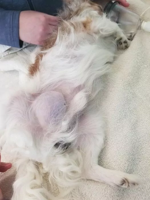
Canine cryptorchidism is the term used to define a medical condition seen in dogs (very rare in cats) in which one or both testicles are retained in the abdomen instead of descending in the scrotal sac. The testicles are a pair of reproductive organs in the male animal. Their role in the reproductive system is to produce sperm and maintain the animal’s reproductive health.
The testicles develop behind the kidneys while the puppy is in utero, and if everything is normal, they will ascend into the scrotum by the time the pup reaches two months of age. In some dog breeds or certain dogs, this ascension might happen later than two months of age. Veterinarians tend to wait until at least six months of age to officially declare a dog a cryptorchid.
The Location of Undescended Dog Testicles
In most cases where a cryptorchid dog is involved, the retained testicle is located either in the abdomen or in the inguinal canal. The inguinal canal is the passage through which the testicle was supposed to descend. In these cases, the testicles get stuck in the canal. There are some cases where the retained testicle is located just under the skin in the inguinal area.
Depending on the location of the retained testicle, the veterinarian might palpate the testicle and locate it during regular physical examination. If the testicle is located deep in the abdomen, this might cause difficulties for palpation.
Diagnosis and Signs of a Cryptorchid Dog
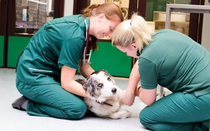
Most experienced veterinarians would agree that if one or both testicles are not physically present in the scrotal sac by two months of age, then that dog is a cryptorchid. It is doubtful that one testicle or both testicles will descend into the scrotum after this age.
The clinical signs and symptoms of a cryptorchid dog are usually not recognized by the animal’s owners. When they buy or adopt the puppy, the testicles are very small, and even veterinarians have difficulties differentiating if one or both testicles are in the scrotum at that time. Young puppies will not show signs of illness with retained testicles unless there is a case of torsion or neoplasia (neoplasias come later in life).
Older cryptorchid dogs, usually older than five years of age, almost always develop neoplasia on the retained testicle. This neoplasia is most commonly a Sertoli cell tumor. This type of tumor originates from the Sertoli cells in the testicle that are a part of the seminiferous tubule. These cells help the process of spermatogenesis.
In older patients, it is indicated to conduct a complete blood count and serum chemistry profile before the surgery. In some cases, the hyperestrogenism caused by the Sertoli cell tumor can cause bone marrow suppression and consequent aplastic anemia.
In older dogs, we should evaluate the biochemistry profile for unrelated chronic diseases such as liver disease or kidney failure. Plain abdominal radiography can be helpful if we suspect testicular torsion or neoplasia.
The retained testicle will be seen on the radiograph as a soft tissue abdominal mass. Chest radiography is recommended to exclude metastasis. Ultrasonography of the prescrotal area, the abdomen, and the inguinal canals can help determine if the tissue is testicular.
Dogs diagnosed with Sertoli cell tumor will show a specific type of clinical signs such as:
- Skin changes (symmetric alopecia)
- The retained testicle will be enlarged and palpable as an abdominal mass
- Feminization syndrome (shrinking of the penis, gynecomastia, taking a female position for urination)
Testicular torsion is another condition seen in cryptorchid dogs. This condition is where the retained testicle will rotate and twist the spermatic cord that brings blood to the testicle. The reduced blood flow will cause swelling and severe acute pain. This condition almost always requires surgical intervention.
Causes and Prevalence of Canine Cryptorchidism
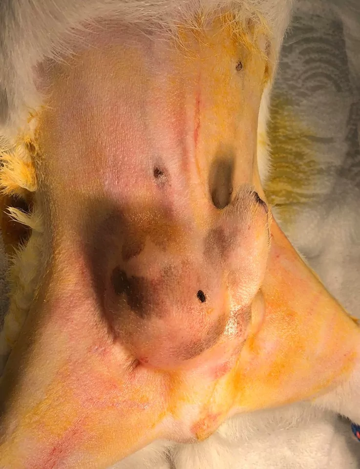
Canine cryptorchidism is a widespread diagnosis in small animal practice. Retained testicles can be unilateral or bilateral. The retained testicle is usually small and/or atrophied, but after a certain age (five years or older) can become enlarged and cancerous.
Canine cryptorchidism is a congenital defect with a reported prevalence of 0.8-10% of dogs. This genetic defect is a sex-linked autosomal recessive trait that is more common in small dog breeds rather than large dog breeds.
The most common dog breeds reported with congenital cryptorchidism are:
- Pomeranians
- Chihuahuas
- Poodles
- Yorkshire terriers
- Maltese dogs
- Miniature Schnauzers
- Siberian husky
- Brachycephalic breeds
Even though cryptorchidism is most commonly seen in the above-mentioned dog breeds, it can also be seen in large dog breeds and mixed breeds.
The sperm develops in the testicles within a system of tubes called seminiferous tubules. For this to be successful and to produce healthy sperm, a specific temperature must be achieved. This is the reason why the testicles are in a scrotal sac outside of the body.
In a bilateral cryptorchid dog, both testicles are inside the body, either in the abdomen or the inguinal canal. This will mean that the testicles will be surrounded by a higher temperature than needed for healthy sperm production, and because of this, that dog will be infertile.
Unilateral cryptorchid dogs are usually fertile but only from the descended testicle. Even though that testicle can produce healthy sperm, it will also pass the gene for cryptorchidism. This means that the healthiest and most ethical solution is the bilateral castration of a cryptorchid dog.
Undescended testicles are 13 times more likely to develop neoplasia and have an increased risk of torsion.
Is Neutering my Dog my Best Option?
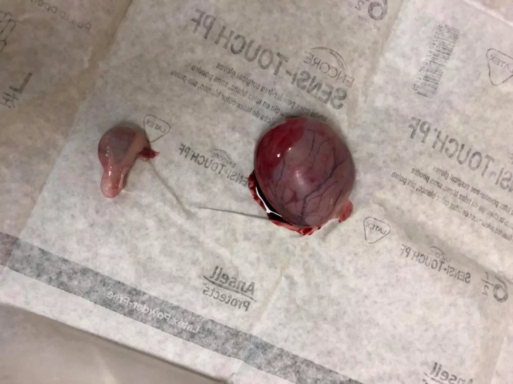
In short, yes!
The first thing you do is remove this genetic defect from the breeding line by neutering a cryptorchid dog. Cryptorchid dogs should never be bred, but some breeders do not care about hereditary conditions and congenital disabilities. This is why you should always get your pet from a reputable breeder.
With neutering a cryptorchid dog, you eliminate the chance for the testicle to develop a tumor or a torsion. Testicular tumors are very aggressive and fast-growing and will cause pain and discomfort to the dog. Testicular torsions are rare but very painful and always require immediate surgical attention.
By neutering a cryptorchid dog, you are increasing his life expectancy. In some countries, there are prosthetic testicles, called neuticles, that can be inserted into the scrotal sac instead of the original testicles. The neuticles have only cosmetic purposes and in no way can replace the real testicles.
The Neutering Process
Because of the nature of the condition and the hereditary factor, surgical removal of both testicles is always recommended. Some dog owners will insist on surgically descending the retained testicle into the scrotum, but this is not a solution to the problem and is not recommended.
The retained testicle(s) can be located either in the prescrotal area, the inguinal canal, or the abdomen.
- Suppose the testicle is in the prescrotal area. In that case, it can be removed either by making an incision directly over the testicle or by pushing it caudally and exposing it through the standard midline incision.
Either approach offers easy exposure of the testicle. The testicle can be removed in an open fashion (incision through the tunic) or a closed fashion (leaving the tunic intact).
- If the testicle is located in the inguinal area, this will require an incision directly over the inguinal canal. Thorough dissection is needed to expose the testicle entirely – the veterinary surgeon must be very careful not to injure the pudendoepigastric artery and vein or any of their branches. It is very easy to confuse the testicle with the inguinal lymph node.
Careful examination and dissection are a must before proceeding with excision. Once the testicle and its structures are exposed, they should be removed as with a normal testicle.
- If the testicle is retained in the abdominal cavity, it can be removed either with a paramedian approach or a ventral midline approach. If both testicles are contained in the abdomen, then this should be approach paramedian.
The paramedian approach allows the advantage not to dissect around the prepuce and the caudal superficial epigastric artery and vein. The paramedian approach avoids creating dead space in the subcutaneous tissue. The formation of seroma is widespread in this area, so it should be expected.
Post Cryptorchidism Surgery Recovery
It is wise to keep all tissue removed and sent for histopathology to ensure that all testicular tissue is removed and examined for neoplasia.
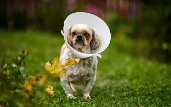
The post-surgical care for a cryptorchid dog closely monitors the incision site for inflammation, possible seroma, or infection. It is advised, no matter how calm or trained, all dogs to wear an Elizabethan collar post-surgery to prevent them from licking the incision.
Resting and easy exercise (just for physiological needs, no running, no mountain climbing, no matter how well the dog feels) is recommended. Analgesic therapy with NSAIDs is recommended usually three-five days after surgery.
Most common post-surgical complications include seromas, incisional dehiscence, ureteral ligation, inadvertent prostatectomy, hemorrhaging due to inadequate ligation of the testicular blood vessels.
Summary
Canine cryptorchidism is a hereditary condition that can be seen in about 10% of purebred dogs. This condition is described as unilateral or bilateral retention of the testicles either in the abdominal cavity, the inguinal canal, or in the prescrotal area under the skin.
Most commonly, small dog breeds are affected but can also be seen in large dog breeds and mixed breed dogs. There is no cure for this condition, and surgical removal of both testicles is recommended. Cryptorchidism in dogs is congenital, and it is recommended that all cryptorchid dogs are removed from the breeding line.
The clinical signs and symptoms of cryptorchidism in dogs can be overlooked by the owners since they do not show any pain or discomfort until late. After a certain age (usually above five years of age), the retained testicle(s) tend to transform into a Sertoli cell tumor. Another complication that is very painful and acute is testicular torsion.
If addressed on time, while the dog is still young and vital, this congenital problem can be fixed by simple neutering. If you suspect that your dog might be a cryptorchid, schedule a visit to your vet and discuss the problem.
