Lymphangiectasia in dogs can be caused by a wide variety of diseases or conditions. Most of these conditions are idiopathic (without a known cause), congenital, or acquired due to a secondary cause.
The most common and frequently encountered secondary cause of lymphangiectasia is Protein Losing Enteropathy. Even though the name sounds scary, lymphangiectasia can be managed effectively when it is detected early.
Lymphangiectasia (lymph [vessels] + angiectasia [dilation]) occurs when the lymph vessels dilate due to an obstruction or inflammation following an infection. It can occur anywhere in the body where there are lymph vessels that drains out lymphatic fluids. Even though it can affect dogs of all breeds, some dog breeds are particularly susceptible.
Defining Protein Losing Enteropathy in Dogs
Diseases can be scary when you first hear of them, but Protein Losing Enteropathy (PLE) can be broken down into four main blocks – Protein Loss Intestine (Entero) Disease (Pathy). It is defined as an intestinal disease where there is the loss of proteins in the intestinal tract through leakages.
There are two major types of proteins in the blood – albumin, and globulin. Albumin is responsible for regulating oncotic pressure in blood vessels and also aid in the transport of many substances through the blood.

Globulin is responsible for fighting infections, blood clotting and serves as transport proteins for iron, copper, and lipids.
Aside from these two proteins, others include coagulation proteins, regulatory proteins, and clotting factors. All these are lost during PLE, but the major and more observable ones are albumin and globulin, as they cause more significant damage when lost.
The Causes of PLE in Canines
The cause of the loss of either one of these proteins or both of them is vital in diagnosing Protein Losing Enteropathy in dogs. PLE can be caused by; Inflammatory Bowel Disease (IBD), a heavy load of parasites in the Gastrointestinal tract, diseases that obstruct the Gastrointestinal tract, bacterial and viral infections like parvovirus enteritis, fungal infection (Histoplasmosis), Crypt disease, “and” some cancers like lymphoma
PLE is most commonly found in dog breeds such as Maltese, Wheaten Terrier (soft-coat), Shar Pei, and Basenji dog breeds.

What is Intestinal Lymphangiectasia in Dogs?
Intestinal lymphangiectasia is a severe form or result of Protein Losing Enteropathy in dogs and the most common type of lymphangiectasia in this species. This condition simply means the lymph vessels in the intestine are dilated.
Lymph vessels are part of the lymphatic system, which carries lymph from the body to the heart, and it is pumped by contraction and relaxation of the body’s muscles. Lymph contains proteins, white blood cells (the majority being lymphocytes), salts, water, “and” fats.
In the gastrointestinal tracts, lymph vessels are called lacteals. These lacteals are designed to absorb fats through the microvilli, which absorb all nutrients.
If these lacteals are blocked or inflamed, they may dilate to make up more room to continue their work. If the pressure is too high, the lacteals burst and release their contents (cells, fats, proteins) in the intestine. The body tries to absorb most of these lost nutrients, but if the cause of the dilation is widespread, most of these nutrients, especially protein, would be lost in the feces.
The causes of lymphangiectasia could be congenital (especially in breeds predisposed to it), idiopathic, and acquired due to secondary disease (mainly from the grounds of PLE).
What are the Symptoms and Signs of Lymphangiectasia?
Your dog may have lymphangiectasia if you notice the following:
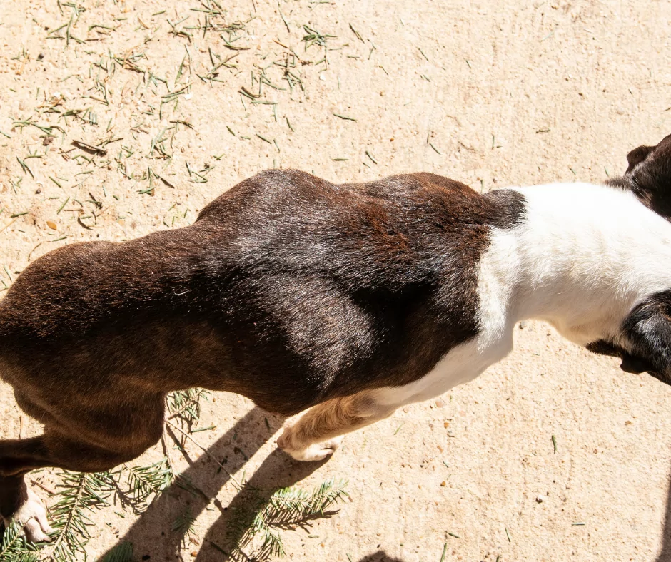
- Weight loss
- Edema (accumulation of fluid in the extremities)
- Ascites (accumulation of fluid in the abdomen)
- Vomiting
- Dehydration
- Respiratory distress is a result of fluid accumulating around the lungs.
- Anorexia

- Lethargy
Always remember that these symptoms are similar for a wide range of diseases, and your dog should be sent to a veterinarian for the proper diagnosis.
How is it Diagnosed?
Lymphangiectasia is diagnosed through a variety of laboratory and clinical examinations. These include:
- Palpation of lymph nodes to test for obstruction.
- Complete Blood Count (CBC) to test for lymphopenia.
- Blood Chemistry to test for low albumin levels, low globulin levels, low calcium levels, low cholesterol levels, and low magnesium levels in the blood.
- Also included in blood chemistry profiles are liver function tests for serum bile acids.
- Renal/glomerular function test which includes urinalysis, urine protein,” and” creatinine ratio are also tested.
- Fecal α1-protease inhibitor assay is used to determine the presence of gastrointestinal protein loss in the feces.
- Other tests for the presence or absence of gastrointestinal diseases.
- Endoscopy to detect intestinal inflammation.
- Biopsies for histological confirmation.
- Abdominal ultrasound to detect fluids around the abdominal organs.
- Thoracic radiographs to detect fluids around the heart and lungs.
Available Treatment Options
There is no known cure for lymphangiectasia. If your dog has been diagnosed with lymphangiectasia, your veterinarian would like to treat the disease’s primary or leading cause first. This is done to manage the disease and provide your dog with an everyday, happy life as much as possible.
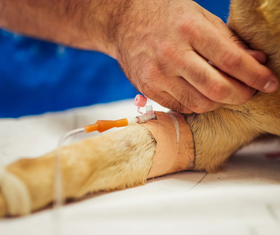
Depending on the underlying cause, your dog might receive parenteral nutrition via IV fluids if it can’t eat and is still vomiting. If your dog has severe coagulation problems due to loss of coagulation protein, your veterinarian will give some plasma therapy to replenish the lost coagulation proteins. Your dog might be put on a special diet to help the body make up for the lost proteins and other nutrients.

Anti-inflammatory drugs (corticosteroids), mainly prednisone, might be prescribed if inflammation is still persistent. Diuretics would also be prescribed to help drain the body fluids. Anticoagulants would also be given to prevent blood clots and embolism from occurring.
A vitamin supplement would also be given to supplement the loss of Vitamin A, B12, D, E, K.
These treatment options depend on the observation made by your Veterinarian, and they might differ from dog to dog even.
How do you Feed Dogs with Lymphangiectasia?
One of the available options for treating a dog with lymphangiectasia is to change the dog’s diet. This new diet, known as the Protein Losing Enteropathy diet, has to be high in protein (20-25% crude protein) to compensate for the high protein loss and low in fat (15% crude fat) to reduce the fat in the intestinal tract. The diet should be highly absorbable to reduce intestinal malabsorption of fats and leakage of lymph.
Home-made diets are often appropriate as the calories, and nutrient levels can be regulated. A Veterinary Nutritionist should be consulted to ensure you are on the right track if you want to prepare home-made diets.
The most common foodstuff and ingredients used to manage lymphangiectasia are; For protein – egg whites, turkey breast, and/or no-fat cottage cheese. For carbohydrates, well-cooked white rice, boiled potatoes without the skin. A small amount of corn oil is added to provide fatty acids for a balanced meal.

A commercial low-fat diet can also be purchased. Always check the calorie label and consult a Veterinary Nutritionist when in doubt. The most common commercial low fat-diets are Royal Canin’s Low Fat and Purina Veterinary Diets H/A.
Summary
Lymphangiectasia, when noticed early, can be managed effectively with dietary changes and supplementary drugs. There’s no fixed cure for the disease unless the underlying conditions are treated, and diet is changed.
Dog breeds that are predisposed to lymphangiectasia don’t mean they would be getting the disease in their lifetime. Still, they have an increased chance of getting lymphangiectasia as compared to other dog breeds.
Life expectancy for dogs with lymphangiectasia and PLE is between five to seven months if it is not treated, managed, or detected early. Your dog can live a happy, healthy long life if your Veterinarian’s instructions are followed carefully.
If you enjoyed this article, you could also read our article on Cystitis in cats.
[simple-author-box]
