
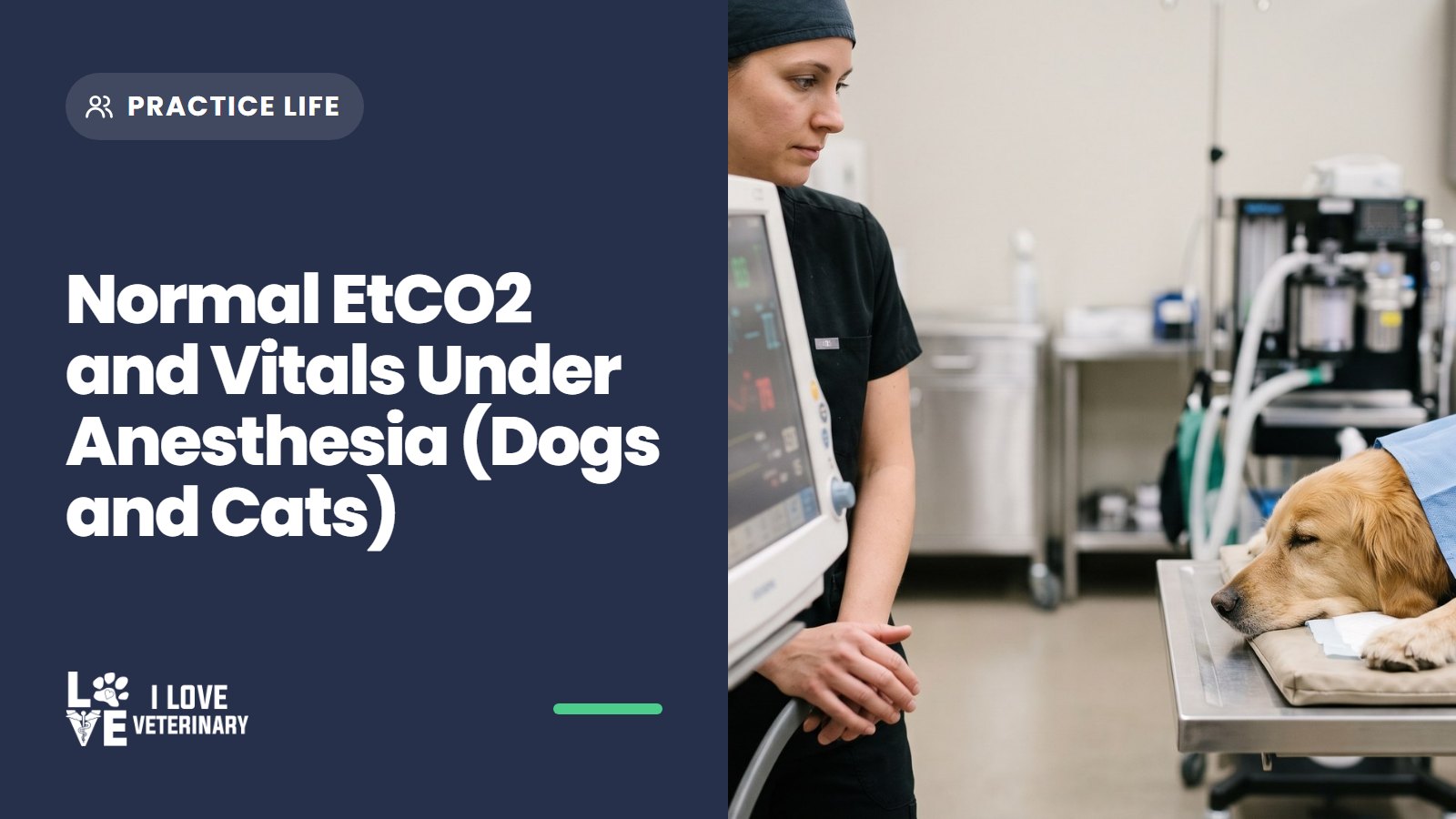
Normal EtCO2 for a dog under anesthesia is about 40-50 mmHg, and up to 55 mmHg is still acceptable in an appropriate surgical plane, according to the 2020 AAHA Anesthesia and Monitoring Guidelines. The 35-45 mmHg you see quoted everywhere is AAHA's range for an awake patient. Above 60 mmHg is hypercapnia, and that is the number that should make you speak up.
The same guidelines cover cats, and they set thresholds for the other numbers on the monitor too. Below is all of it in one place, what each monitor is really telling you, and when to tell the anesthetist or the vet. For awake values (the ones you take in the exam room), use our chart of normal vital signs in dogs and cats.
AAHA thresholds at a glance (dogs and cats)
One thing to know before you read the table: AAHA gives complication thresholds, not a "normal range" for every parameter. Where it gives no number, we say so rather than borrow one.
Parameter | What AAHA says | Tell the anesthetist or vet when |
|---|---|---|
EtCO2 (end-tidal CO2) | About 35-45 mmHg awake; about 40-50 (up to 55) mmHg in an appropriate surgical plane | EtCO2 is above 60 mmHg (hypercapnia) |
SpO2 (pulse oximetry) | Hypoxemia is SpO2 below 95%; severe below 90% | SpO2 is below 95% |
Blood pressure | Hypotension: systolic below 80-90 mmHg, mean (MAP) below 60-70 mmHg, diastolic below 40 mmHg. Hypertension: MAP above 120-140 mmHg or systolic above 160-180 mmHg (uncommon under anesthesia) | Pressure crosses either line |
Heart rate, too fast | Tachycardia: cats above 180 bpm; dogs above 150 bpm (large) to 190 bpm (small) | Heart rate passes the line for that patient |
Heart rate, too slow | AAHA gives no number for bradycardia | The rate falls or the rhythm changes (the number is set with your anesthetist per patient) |
Respiratory rate | AAHA gives no normal number under anesthesia; judging rate and depth is "very subjective" | Breathing changes; look at EtCO2 to see what it means |
Body temperature | Hypothermia is core body temperature below 98°F (36.7°C) | Temperature is below 98°F (36.7°C) |
Gum color | "Not a sensitive indicator" of oxygen: cyanosis appears only when hypoxemia is profound | Gums look pale, gray or blue (but don't wait for this) |
Source: Grubb T, et al. 2020 AAHA Anesthesia and Monitoring Guidelines for Dogs and Cats. Thresholds apply to dogs and cats unless the row says otherwise.

AAHA's list of what to watch is respiratory rate, SpO2, EtCO2, blood pressure, heart rate and rhythm (ECG), capillary refill time, mucous membrane color and body temperature, plus hands-on checks by the anesthetist. Shorthand on the monitor tripping you up? Our list of veterinary abbreviations covers the common ones.
What each monitor tells you (and how it can fool you)
AAHA wants decisions to come from both the electronic monitors and the anesthetist's own assessment of the patient. Most complications show up on a monitor first, but monitors also produce readings that aren't real. Knowing the usual artifacts saves time, but it never replaces telling someone.
Capnograph (EtCO2)
EtCO2 is the carbon dioxide in the breath at the end of each exhalation. It shows, without a needle, how well the patient is ventilating, and it usually runs a few mmHg below the arterial value (PaCO2). Changes in EtCO2 tend to show up before SpO2 falls.

The waveform matters as much as the number. A capnograph shows apnea and airway problems quickly, and the adapter on the endotracheal tube can confirm the tube is in the trachea during intubation.
Common artifacts and look-alikes:
- Sidestream line kinked, clogged with secretions or wet: readings go wrong or disappear. Sidestream monitors also show the value a few seconds late.
- Cuff leak: CO2 escapes around an underinflated tube cuff. You may hear a hiss on expiration or smell inhalant near the mouth.
- Obstruction: a kinked or blocked endotracheal tube, or a blocked expiratory limb of the circuit, changes the waveform shape.
- Rebreathing: the baseline no longer returns to zero between breaths. Exhausted CO2 absorbent or a faulty one-way valve can cause it.
- Mask readings: a sensor on a face mask is less accurate than one on an endotracheal tube.
Tell the anesthetist when EtCO2 climbs toward 60 mmHg, when the waveform changes shape, or when it goes flat.
Pulse oximeter (SpO2)
SpO2 is the percentage of hemoglobin carrying oxygen. AAHA calls pulse oximetry the best way to assess oxygenation continuously, and hypoxemia is uncommon in an intubated patient breathing oxygen.
The trap is blaming the probe. AAHA's order of checks starts with the patient, not the clip: is the patient properly intubated, connected to the oxygen source, with an adequate oxygen supply? Poor perfusion can lower the reading too, so a low SpO2 alongside a slow capillary refill, weak pulses or low blood pressure is not a probe problem.
So say the reading out loud first, then let the anesthetist work through those checks with you.
Blood pressure
AAHA lists hypotension among the most common anesthetic complications. Capillary refill time and pulse quality help confirm poor blood flow.
High readings are uncommon under anesthesia. Before anyone trusts one, AAHA suggests checking that the cuff hasn't slipped down the leg (toward the carpus, for example) and confirming the pressure with a second method such as Doppler. For the awake picture, see normal blood pressure in dogs and cats.
ECG and heart rate
AAHA's point about a fast heart rate is worth remembering: it should start a list of possible causes, not an assumption that the patient is too light. Pain or surgical stimulation, low oxygen, high CO2 and low blood volume are all on that list. Sorting them out belongs to the anesthetist and the vet; your job is to report the rate and what was happening at the time.
Rhythm problems such as sinus tachycardia, sinus bradycardia, AV block and ventricular arrhythmias are watched with auscultation or ECG. A mismatch between the pulse and the heart rate can also show up on a Doppler or the SpO2 waveform.
There is no AAHA number for bradycardia. The line is set with your anesthetist per patient, so report a falling rate early.
Respiratory rate
The honest answer is an unsatisfying one: AAHA gives no normal respiratory rate for a dog or cat under anesthesia. Judging rate and depth by eye is "very subjective", in AAHA's words, and a patient can breathe at a reasonable-looking rate and still retain CO2. The capnograph, not the count, tells you whether ventilation is adequate.
Count breaths anyway and record them. A change in rate is a reason to look at EtCO2.
Temperature
Hypothermia slows drug metabolism, causes heart and breathing problems, leads to poor wound healing and can drag out recovery. AAHA wants warming started early and continued through recovery, so temperature belongs on the record from the start of the procedure, not just at the end.
Hands-on checks
AAHA describes a surgical plane as absent palpebral reflex, mild jaw tone and no purposeful movement. Gum color and capillary refill time add context, with one warning: by the time gums look blue, hypoxemia is already profound.
When to tell the anesthetist or vet
AAHA names the most common complications as hypotension, hypoventilation, hypoxemia, hypothermia and some arrhythmias. Any line in the table above is a reason to speak up right away. So are these, which have no number attached:
- the capnogram goes flat or changes shape
- the heart rate falls or the rhythm changes
- breathing changes in rate or depth
- the patient moves, or reflexes return

Check for the obvious artifact while you say it, not before. A probe that fell off takes seconds to spot; a real drop in oxygen doesn't wait for you to finish troubleshooting.
Nearly half of dog and most cat anesthetic deaths happen in recovery
The 2020 AAHA Anesthesia and Monitoring Guidelines report that 47% of anesthetic-related deaths in dogs and 60% in cats happen after the procedure, mostly within the first 3 hours.
Breathing often stays depressed in early recovery, with high CO2 and low oxygen. AAHA's guidance is to keep the capnograph adapter on the tube until extubation, use a pulse oximeter throughout recovery, and keep monitoring heart rate, respiratory rate, SpO2, blood pressure and temperature.
The finish line: keep watching until the pet is alert, warm and walking.
Keeping the anesthesia record
AAHA lists documentation of patient parameters during anesthesia and recovery as one of the actions that lower anesthetic risk. A good record:
- logs every monitored parameter through the procedure and into recovery, so a trend is visible before it becomes a crisis
- notes previous anesthetic events and recoveries, which AAHA says should be checked before the next anesthetic
- documents the pre-anesthetic physical exam (AAHA cites a study where failing to record a physical exam increased the odds of death in dogs)
- goes alongside an anesthesia safety checklist, which AAHA recommends
AAHA also notes that most complications are first picked up on an anesthetic monitor, so whoever keeps the record should be comfortable reading ECG, EtCO2, blood pressure, SpO2 and temperature. That skill sits at the heart of what vet techs do on the surgery floor, and if it's the part of the job you love, our guide to veterinary technician specialties shows where a credential can take it.
Sources
- AAHA: 2020 AAHA Anesthesia and Monitoring Guidelines for Dogs and Cats (Grubb T, et al., JAAHA 2020;56(2):59-82)
- dvm360: Ventilation and ETCO2, it's more than just breathing (Proceedings)
- Synergy CPD: Capnography, getting started (Claire Roberts RVN)
This article is for general information and is not a substitute for advice from your veterinarian.